植物抗病机制示意图可直观呈现信号通路与分子互作,助力解析抗病机理,为抗病育种、靶点筛选及作物病害绿色防控提供理论支撑,另外,漂亮的机制图也能让审稿人和读者觉得文章作者更加用心!
(Nature Communications,2026)
那么,如何轻松绘制上图这般好看的植物抗病机制示意图?这里为大家推荐一个在线Ai绘图工具!
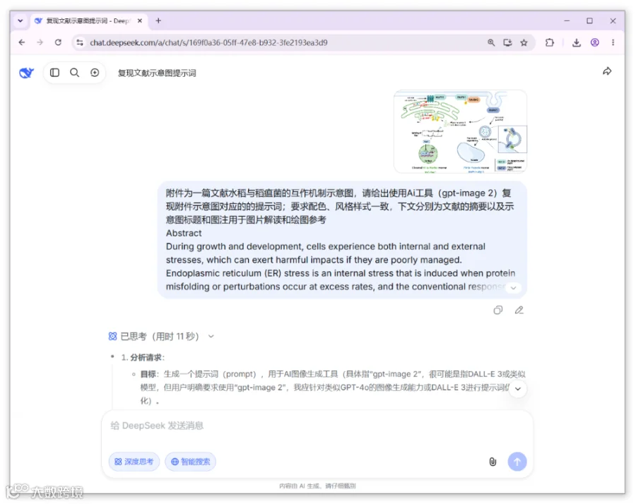
对于上图植物抗病机制示意图的绘制,这里以一篇标题为 “Magnaporthe oryzae MoPh1 perceives ER stress and promotes adaptive responses via a plasma membrane-to-vacuole pathway” NC文章中的示意图为例。对于绘图提示词的准备,我这里直接使用DeepSeek进行生成。这里将原文的示意图上传为附件,并直接复制文献的摘要、示意图标题和图注用于图片解读和绘图参考!
附件为一篇文献水稻与稻瘟菌的互作机制示意图,请给出使用Ai工具(gpt-image 2)复现附件示意图对应的的提示词;要求配色、风格样式一致,下文分别为文献的摘要以及示意图标题和图注用于图片解读和绘图参考
During growth and development, cells experience both internal and external stresses, which can exert harmful impacts if they are poorly managed. Endoplasmic reticulum (ER) stress is an internal stress that is induced when protein misfolding or perturbations occur at excess rates, and the conventional response pathways from the ER to the nucleus are activated to address the stress. However, the involvement of the plasma membrane (PM) system in response to this internal stress has been insufficiently investigated. Here, a PM sensor, MoPh1, was observed to perceive stress through ER-PM contact sites and target the autophagosome and vacuole, consequently stimulating the autophagy process and supporting stress relief. The PM-to-vacuole pathway mediated by MoPh1 is independent of the classical ER-to-nucleus pathway and might be highly important in both fungi and plants, as it plays a crucial role in alleviating ER stress and promoting cellular adaptation for cell survival.
Fig. 7: Model of the PM-to-vacuole response manner under ER stress.The transmission of ER stress signal to the PM sensor MoPh1 occurs via EPCSs during the early stage of stress response, and then induces MoPh1 phosphorylation by the protein kinase MoDbf2, followed by targeting autophagosomes, which facilitates the formation of the autophagosome and its consequent fusion with the membrane vesicles to promote autophagy and thus help alleviate the stress. This stress response from PM-to-vacuole could also promote the formation of MoIre1 phase-separated clusters for manufacturing the efficient MoIre1 machinery and the activation of the classical ER stress response, which are fundamental to ER stress response and enhancing the adaptation of both fungi and plants to this stress.
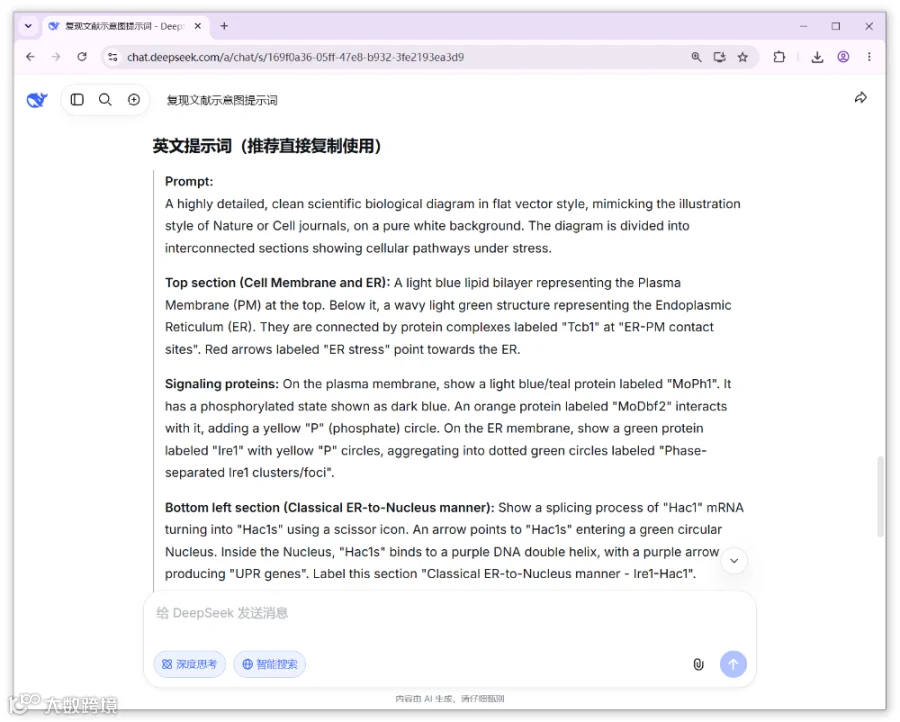
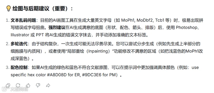
经过11秒的深度思考,包括解构图像与科学内容、生成提示词、润色提示词等步骤,DeekSeek很快给出了中英文提示词,如下图,并给出了绘图与后期建议。
在Ai绘图工具页面,图片类型参数选择图片摘要,模板类型参数选择生物系统机制图,然后直接复制DeepSeek给出的提示词,如下图。
https://www.omicshare.com/tools/home/report/report_ai_image.html
点击提交按钮后,在页面下方结果展示板块,可查看初始的绘制效果,如下图。
初始绘图效果基本满足需求,点击下载图片按钮,图片会自动保存为png格式,绘图如下:
此外,我们也可以切换参数,试试其他模板类型,例如,点击填充当前任务参数按钮,将模板类型调整为CellPress期刊风格图,重新提交绘图任务,绘图效果如下。
当然,同样的方法我们也可以重新使用豆包生成提示词,再次复制到Ai绘图工具中,模板类型继续选择生物系统机制图生成示意图。
另外,也可以继续点击填充当前任务参数按钮,将模板类型调整为CellPress期刊风格图,绘图效果如下图,也是很好看的!
好啦,本次的植物抗病机制示意图绘制教程就分享到这里啦!
https://www.nature.com/articles/s41467-026-70610-0#Abs1
随着技术成本下降与分析算法迭代,单细胞引领的多组学整合方案,必将成为癌症等疾病基础研究与临床转化的标配工具。如果您有项目需求,欢迎联系基迪奥生物,我们将为您提供定制化的项目解决方案,一起冲击顶刊。
*未经许可,不得以任何方式复制或抄袭本篇文章之部分或全部内容。版权所有,侵权必究。
联系方式:020-39341079;service@genedenovo.com
测序未来,从加入基迪奥开始
识别二维码,了解岗位,开启事业新篇章
#单细胞转录组#多组学#肠道炎症#肠上皮#文献解读





 SCIPainter