

What is cartilage?
Cartilage is a very strong, smooth, elastic, fibrous structure found in joints and at the end of long bones. In the body it takes many forms and has multiple purposes throughout the body.
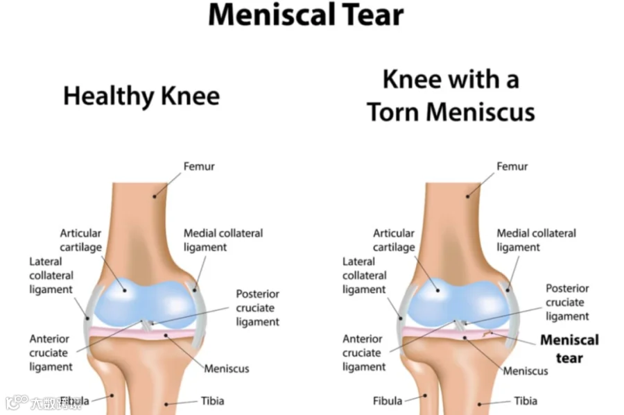
In the knee we have two types of cartilage; articular cartilage and the menisci cartilage.
The articular cartilage is located on the end of the bones; the femur (above the knee) and tibia (below the knee). Articular cartilage is involved in osteoarthritis.
The menisci are situated in between the bones, on top or below the articular cartilage, they are not connected to the articular cartilage.
For this article we are going to solely focus on menisci cartilage.
What is the function of meniscus?
The menisci are a fibrocartilaginous structure that provide the joint with increased stability, distributes body weight evenly through the knee and increases shock absorption.
There are two menisci. The one on the outside, also known as the lateral meniscus and the one on the inside, known as the medial meniscus (see image below). The medial meniscus is the most commonly injured of the two.
Each meniscus has an anterior and posterior horn and most tears occur at the posterior horn.
A torn meniscus is a common injury that can affect active and sedentary patients of all ages.
How do you tear the meniscus?
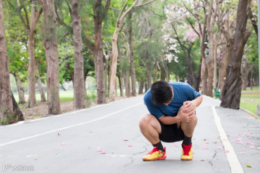
The meniscus is most commonly injured by a twisting or turning movement whilst your foot is planted on the floor. In a young sporty person this could be due to turning quickly when playing a game of football or a tackle during a rugby match.
The effect of this twisting movement puts torsion throughout the meniscus and causes the fibres to tear. This results in an acute tear of the meniscus. Some people report they hear their knee click and feel pain immediately.
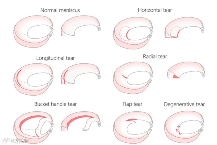
There are many different types of tears (see image below). The different tears can present with their own specific symptoms. For example, a ‘bucket handle tear’ may cause ‘locking’ of the knee. ‘Locking’ is when the knee gets stuck in one position and you cannot move it. This is very painful and debilitating when it happens. Not everyone with this type of tear experiences this problem.
As we get older meniscal tears can occur with the same twisting mechanism but they can also occur due to seemingly innocuous movements such as misjudging a step or turning whilst walking. It is not unusual that there is no specific incident or trauma, before the knee becomes painful.
How do you know if you have a meniscal tear?
The symptoms can vary from person to person but the key signs and symptoms are:
· Swelling – this normally takes 24 hours to develop after the onset of pain or injury
· Tenderness on the inside (medial meniscus tear) or outside (lateral meniscus tear) of the knee
· Pain with straightening the knee
· Pain and an inability to bend the knee fully i.e. unable to pull your heel into your buttock
· ‘Locking’ and ‘clunking’ of the knee
· Pain whilst squatting, twisting and kneeling
How is a meniscal tear diagnosed?
Meniscal tears are quite straightforward to diagnose. Clinical assessment has been shown to be around 80% accurate for diagnosis of meniscus tears. Your physiotherapist will carry out specific clinical tests to assess for tenderness on the joint line (where the meniscus is located), your range of movement, the amount of swelling in your joint and the stability of your knee. There is a specific meniscal test called the ‘McMurray’s test’ that your physiotherapist may additionally carry out to help establish your diagnosis.
If we are unsure of your diagnosis or we would like to understand more about the extent and type of the meniscal tear then we may refer you for an MRI scan. MRI scans are also very useful to assess for any other contributing factors such as an associated ligament tear. We can refer you directly without going to your GP.
Some of our clinicians are also fully qualified musculoskeletal sonographers and will carry out a diagnostic ultrasound scan of your knee.
Ultrasound is unable to assess the deep portion of the meniscus, however, it is an effective tool to assess for any swelling and inflammation in the knee. Ultrasound can also visualise meniscal cysts and other structures in the knee such as the medial and lateral collateral ligaments and the surrounding tendons. Meniscal cysts are small fluid-filled pockets that arise from the meniscus. These can occur secondary to a tear.
Will your meniscus heal?
This is not a simple yes or no answer! There are many factors that will determine whether a meniscal tear will heal or not. Age and location of the tear are two of the main variables. Even if the tear does heal it often takes months rather than weeks to recover. The blood supply to the meniscus is poor and so any healing is relatively slow, compared to a muscle for example, which has a rich blood supply.
The younger you are, the more likely it is to heal. Unfortunately over the age of 30, meniscus tears are far less likely to heal. The good news is most of these tears even though they do not heal, become pain free. This is supported by a significant body of evidence that patients with a meniscus tear in their knee observed on MRI, are often asymptomatic. Like many places in the body e.g. the lower back, there is a poor correlation between structural abnormalities and levels of pain.
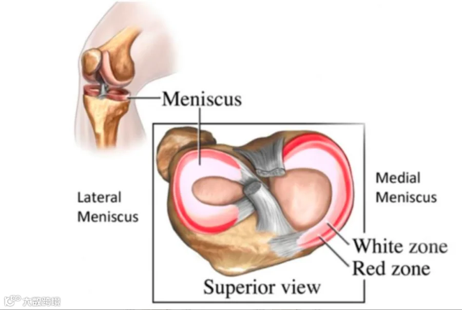
The location of tear can influence whether a meniscal tear will heal or not. As mentioned earlier the menisci have a relatively poor blood supply. The blood supply varies depending on the location of the tear. It can be split into a white zone and a red zone (see image below). The red zone is located on the periphery (the outside) of the meniscus and has superior blood supply to the white zone, which is located in the deeper portion. If a tear is located in the red zone, they have more potential to heal. To determine exactly where your tear is located you would require an MRI scan.
How do you treat a torn meniscus?
Gaining a correct diagnosis is of paramount importance and is the first stage of getting back on the road to recovery. Your age, mechanism of injury, location, type and size of tear will influence which treatment is required. But it is not as simple as this, as patients can present very differently with the same injury.
At Complete Physio each patient is treated as an individual.
Following a comprehensive assessment your physiotherapist will present the best treatment options available to you. Most clients we see with a meniscal tear do improve with a course of physiotherapy, surgery is rare.
The best solution for you will vary depending on your own individual circumstances. For example, a professional footballer is more likely to have surgery than a sedentary individual or recreational runner, as they do not have the time to ‘wait and see’ whether it heals and/or the pain resolves.
This article is excerpted from:CompletePhysio.
https://complete-physio.co.uk/meniscal-tear/
The copyright belongs to the original author, edited and published by JK-Clive Pain Clinic specializes


JK-Clive Pain Clinic specializes in management of acute and chronic pain caused by various "sports injuries" and "degenerative diseases",we are located in Beijing Sanfine International Hospital and our services can be paid directly by most of the international insurance.


Jingbin Zhou, MD, PhD, Professor.
Board member of Chinese Society of Sports Medicine(CSSM)
Vice Secretary-general of Chinese Association of Sports Medicine(CASM)
Vice Chairman of Youth Committee of Chinese Society of Sports Medicine
Committee member of Asian Athletic Association
Committee member of Chinese Football Association (CFA)
2009 Impuls Rehabilitation Center and Krankenhaus Sports Medicine Hospital, Germany
2010-2011 Orthopedic Department of University of Pittsburgh of Medicine
Center (UPMC) 2011 Hospital for Special Surgery (HSS), USA
Clive Chen
Attending neurosurgeon of Taichung Veterans General Hospital
Neurosurgeon Board and Pain Physician Board
Graduate of Medical School of National Taiwan University
Research Scholar specializing in neuromodulation and pain management, UCLA
Taiwan Pain Society Distinguished Service Award 2015
Feng Lei
Attending physician of JK-Clive Pain Clinic
Graduated from the Department of Medicine, Peking University, MD
Attending physician of Beijing Ji shui tan hospital pain management department
Xu Hao
Orthopaedic specialist, sports specialist
Committee member of Physical Therapy Group of Physical Rehabilitation,
Physical Therapy Special Committee of Chinese Medical Association of Rehabilitation
Visiting scholar, University of Southern California, USA
Visiting scholar, Georgia State University, USA
Member of the National Team of Figure Skating and Freestyle Skiing Aerials Rehabilitation Support Expert Group
Member of National Team Doctor Training Class Lecturer Group


Past recommendations


Light up sharing, like, watching
Look who's watching with you
