
点击公众号即可订阅

State-of-the-Art microCT Imaging
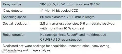
X-ray micro-computed tomography (microCT) is one of the most advanced methods for gaining 3D insights into samples of any material and shape non-destructively, with little to no sample preparation. Bruker microCT, a pioneer of microCT, has now made this technology easier and more accessible for everyone to analyze:
• Biomechanical reactions
• Biomaterials and implants
• Enamel and bone mineralization
• Marginal fit in restorations
• Pulp pathosis
• Root canal morphology
• Skeletal development
• Tissue engineering
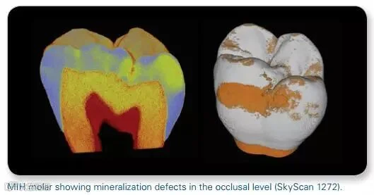
Molar Incisor Hypomineralization (MIH)
• Detect hypomineralized enamel areas
• Measure mineral density defects and distribution
• Make virtual representations of crown mineralization by surface and volume rendering in real time
Periodontics & Orthodontics
• Evaluate tooth movement and root resorption
• Evaluate the micro-leakage at interface of bone and the root
• Dynamic monitoring of bone quality in vivo over time using fast and low dose scans
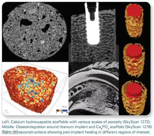
Implants and Scaffolds
• Resolve internal fine structures with 3D non-destructive imaging
• Analysis of osteointegration
• Quantify scaffold porosity (open, closed, interactions, accessibility to bone cells)
• 4D in situ examination under controlled temperature, compression or tension
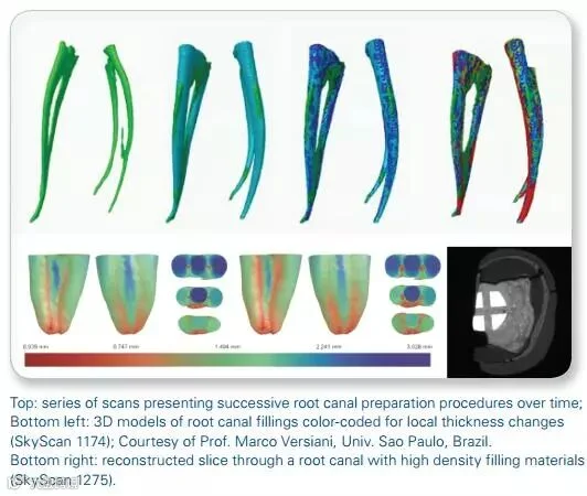
Endodontics
• 4D analysis of the root canal morphology
• Evaluate root canal treatments
• Compare various endodontic instruments and filling materials
• Quantify micro-defects and changes inside root canals
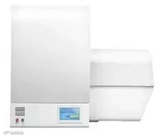
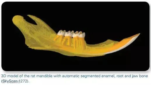
SkyScan 1272
High resolution ex vivo microCT
SkyScan 1275
High throughput ex vivo microCT
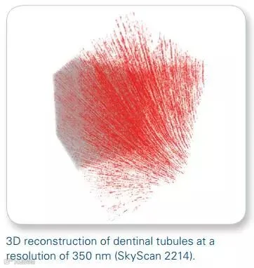
SkyScan 2214
High resolution multiscale nanoCT
SkyScan 1276
High resolution in vivo microCT