
点击公众号即可订阅
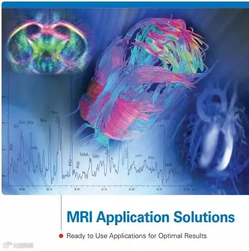
The Bruker Philosophy for Preclinical Imaging
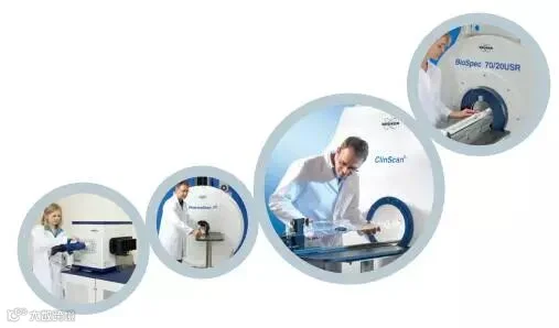
For preclinical MRI Bruker has embraced a unique philosophy to ensure that we understand our customers’ needs and the challenges they face, and that best utilizes our expertise and knowledge. To deliver on this approach Bruker has installed, in-house, eight installed preclinical MRI systems of different bore sizes and field strengths from 1 to 15 Tesla (T) at our main application facility in Ettlingen, Germany.
Supported by many application specialists that cover every application, their trusted expertise and knowledge is what drives the development of innovative in vivo imaging applications. The resulting solutions benefit a wide range of demanding needs in preclinical imaging, molecular medicine, biomedical and pharmaceutical research. It is this same knowledge and expertise that is made available to individual projects and collaborative developmental efforts.
By developing MRI instrumentation in-house on in vivo subjects, the number of unnecessary animal experiments at customer sites is significantly reduced, as is the time required before customers can produce their own data.
The Applications Center has fully optimized protocols for fMRI, DTI, Perfusion, Cardiology,Angiography, Abdomen, Anatomy, IntraGate (Self-Gating), Relaxometry, and Spectroscopy – all of which have been developed in vivo in-house, for use with mice and rats across our entire MRI product range, namely BioSpec®, PharmaScan®, ClinScan® and Icon. These protocols have been developed for all field strengths from 1 T to 15.2 T with the appropriate gradient and RF coils. There are more than 600 protocols available that can be used ‘out of the box’.
Traditionally such powerful technology required years of expertise, intensive training or specialist operators, but today an MRI system from Bruker does all the complex work for you. You focus on your biological,pharmaceutical and preclinical investigations - we’ll take care of the rest.
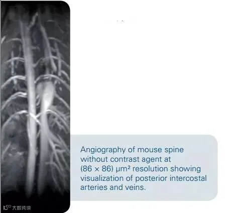
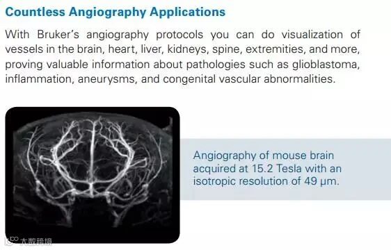
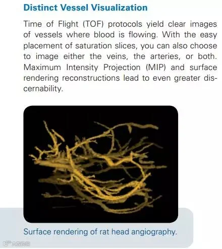
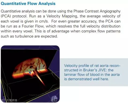
Angiography
Examine the finest vessels with Bruker’s Time of Flight (TOF) and Phase Contrast Angiography(PCA)
• No need for contrast agents
• For all of your pathological questions, all throughout the body
• Qualitative and quantitative analysis
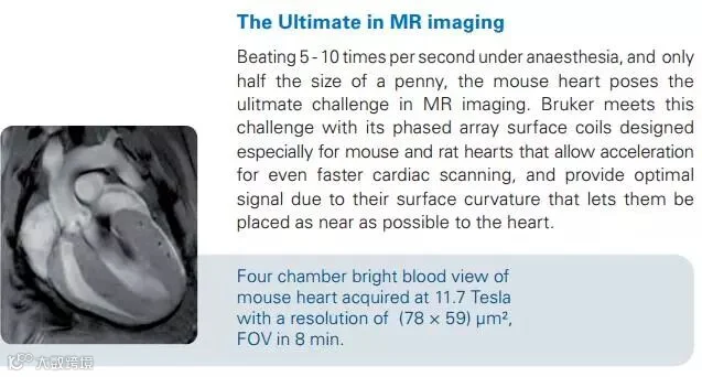
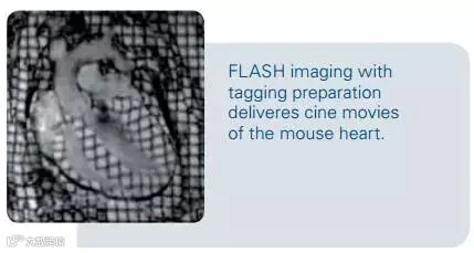
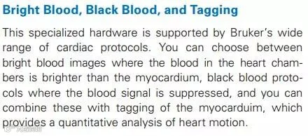
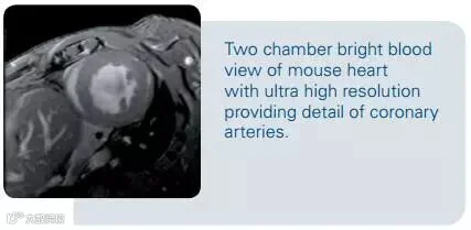
Cardiology
Bruker meets the ultimate challenge in MR Imaging with its unsurpassed cardiac imaging
• Four chamber black blood to locate defects of the septum
• Two chamber bright blood for investigation of ejection fraction
• Short axis tagging for assesment of wall motion
Diffusion
Achieve highest resolution and best possible sensitivity with Bruker’s diffusion options
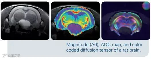
• Measure diffusion weighted images, diffusion tensor images, trace images,fractional anisotropy images,and apparent diffusion coefficient maps
• Acquire highest possible sensitivity in diffusion weighted images with Bruker’s world-wide strongest gradients
• Obtain critical information about tumor infiltration, cardiac infarction,connectivity, stroke, and more
View Diffusion in More than Just One Way
Bruker’s diffusion imaging is more than Diffusion Weighted Images (DWI) and Diffusion Tensor Images (DTI). When you use Bruker’s pre-prepared DWI and DTI protocols, no separate scanning is necessary to receive a non-diffused
A0 image and to calculate trace images, Fractional Anisotropy (FA) images, and Apparent Diffusion Coefficient (ADC) maps. This additional information complements DWI and DTI, since for example, ADC, which measures the magnitude of diffusion, is reduce by approximately 50% minutes after the onset of ischemia.
The Best Gradients for the Best Diffusion Weighted Images
The contrast in Diffusion Weighted Imaging (DWI) originates from the difference in amount of diffusion. Regions that have pathologically disturbed diffusion, such as found when multiple sclerosis, epilepsy, and schizophrenia, stroke, or tumors are present, are easily visible. Greatest sensitivity is achieved with higher b values, which can only be realized with extremely strong gradients. This is just one more reason why Bruker invests so strongly in its world-wide leading gradient technology.
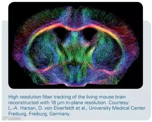
Diffusion Tensor Imaging for Unsurpassed Accuracy
Diffusion Tensor Imaging (DTI) surpasses other imaging methods in addressing many biological questions. Since DTI visualizes the diffusion orientation, it is used to assess fiber connectivity in the embryonic development of transgenic models. It is also the only method that can accurately visualize the level of tumor infiltration into healthy tissue.
fMRI
Where are they thinking? Find out with Bruker’s fMRI
• From visual stimulation to conditioned taste aversion
• The speed you need for fMRI
• Integrated evaluation tool
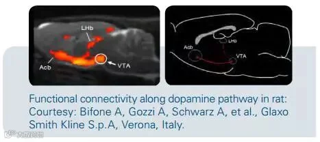
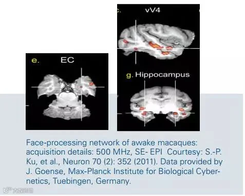
Exciting Possibilities Become Reality
The exciting possibilities of fMRI become reality with the research that Bruker’s customers carry out on a daily basis. Their studies extend beyond forepaw stimulation to discoveries in areas such as face recognition and functional connectivity.
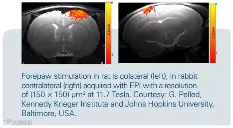
Exceedingly Fast Sequences
Mice and rats, macaques and rabbits are all used and Bruker’s fastest EPI sequences deliver the needed speed by recording images every second. To make this possible Bruker has pushed physics to its extremes by including all
of the mathematical tricks into the software and using its world class coil knowledge when building phased array coils.
Integrated Evaluation Tool
The excitement peaks when the data is put into ParaVision’s integrated FUN evaluation tool and a perfect time course is seen. FUN tool even allows the evaluation of more than one type of stimulation within a routine.
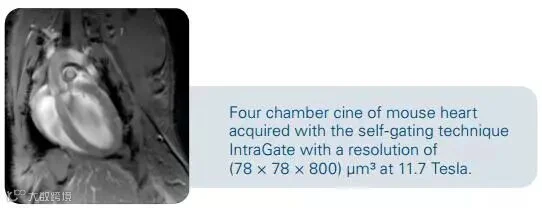
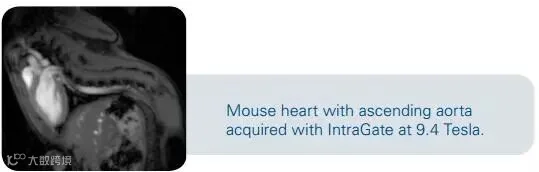
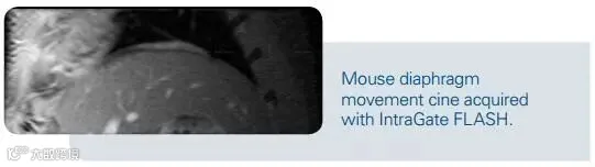
Self-Gating with IntraGate
Bruker’s IntraGate is the electrode-and trigger-free method of scanning the heart and abdomen to assess morphology and disease
• Scans quality as crisp as traditional triggered scans
• Retrospective gating of rapid heart beating and strong respiratory motion in small animals
• Choose between abdominal and bright or black blood multislice cardiac cines
• Choose between cardiac, respiratory, or cardiac in combination with respiratory cines
Electrode-free Cardiac Imaging Eases Setup and Saves Time
Bruker’s patented IntraGate provides artifact-free cardiac imaging without the need for tedious electrode setups. When time is not on your side, you can shorten your animal setup by skipping the electrodes. This is of special interest to users who run their scanners in a “conveyor belt” style, since this critical bottleneck setup time is decreased, allowing a higher throughput. IntraGate is especially useful for animals with particular pathologies that hinder clear and strong electrode signal reception, where triggering is difficult.
No Triggering Equals Worry-free Scanning
IntraGate can be used for cardiac, respiratory, and abdominal imaging, all of which normally require triggering. With Bruker’s IntraGate, there is no need to consider triggering: just select either a time frame cine for abdominal imaging, or a black or bright blood cine for cardiac imaging, set your number of slices, press the traffic light button and sit back and relax while IntraGate takes care of the rest.
Scan Once, Get up to Three Cines
Since Bruker’s IntraGate uses retrospective triggering, you can decide after the scan, what type of cine and even how many frames you would like to have. Cardiac, respiratory, as well as cardiac in combination with respiratory cines are possible, and the number of cine frames can be increased as much as desired, as long as the SNR suffices.
Perfusion
With Bruker you have the full range of perfusion options: dynamic contrast enhanced and dynamic susceptibility contrast studies using contrast agents and contrast agent-free arterial spin labeling
• Cover all of the pathologies that are of interest to you – stroke, tumor diseases, vessel stenosis …
• Blood flow and mean transit time are easily calculated
• Extremely short scan times give you excellent temporal resolution for your contrast agent studies
All Perfusion Options at Your Hand
Bruker offers the full range of perfusion study options: whether with or
without contrast agent, in the brain or the kidneys, you have the choice.
Tumor detection in brain, thorax, and abdomen, tumor neoangiogenesis,
tumor vascularisation, cerebral ischemia, disruption of the blood brain
barrier, vessel stenosis, flow rates, hypervascularisation, and infectious
or inflammatory disease analyses are all possible.
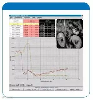
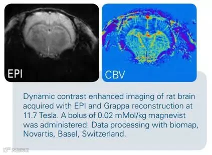
Dynamic Contrast Enhanced (DCE) and Dynamic Susceptibility Contrast (DSC) Studies
A wealth of information can be obtained using contrast agents. Depending on the application, the contrast agent can increase the MR signal, as in tumor neoangiogenesis, or it can decrease the signal, as in stroke, where the amount of perfusion indicates which areas are in the penumbra and which are in the ischemia. For the highest time resolution when using contrast agents, choose Bruker’s fastest sequences, which record an image every two seconds.
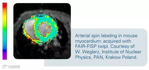
Blood Flow Quantifcation with Arterial Spin Labeling
For calculation of blood flow, no contrast agent is necessary. Bruker’s Arterial Spin Labeling (ASL) imaging sequences come with easy to use quantification software that provides blood flow maps of either the entire scanned area or your desired region with just a few simple clicks. Blood flow values can be read out for each individual pixel.
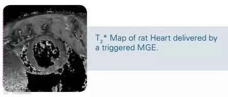
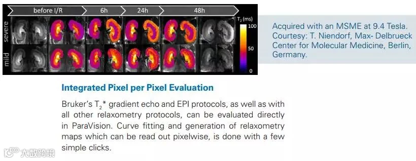
Relaxometry
Gain valuable knowledge about pathologies, contrast agents, and more with Bruker’s
• Relaxometry protocols T1, T2, and T2* maps in stand-alone, combined, or fast variations
• For mapping of all pathologies in all tissues
• Complete analysis directly in ParaVision
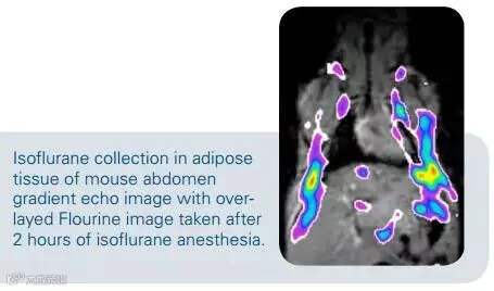
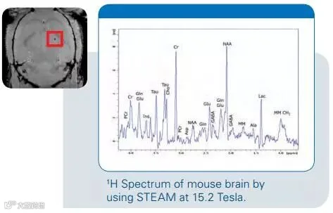
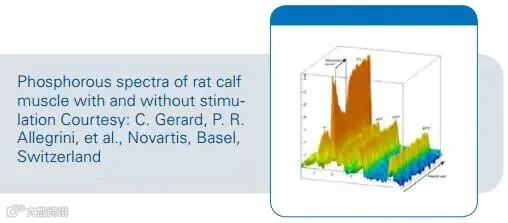
Spectroscopy
• Observe metabolic disorders involving chemicals with only millimol concentrations
• Integrate, filter, and phase your spectra directly within TopSpin
• Perform carbon, fluorine, or phosphorous studies
More than Water
The brain, liver, and muscles of your animals contain more than just water, and MR spectroscopy makes non-invasive studies of metabolic processes in these tissues possible. You can identify metabolic disorders and observe long term changes in metabolic processes even though the chemicals that are detected here are only found in vivo in millimolar concentrations.
More than Single Voxels
You can choose to look at single voxels or perform chemical shift imaging, knowing that Bruker has powerful integrated software for the analysis of
both. This allows you to display multiple single voxel spectra simultaneously, calculate line widths and integrals of your peaks, or overlay a spectral map from your chemical shift image onto a reference image.
More than Protons
With Bruker’s standard carbon, fluorine, and phosphorous coils, glucose uptake, changes in ATP, or effects of isoflurane, can be monitored with little to no background signal, and sensitivity can be increased even further by using Bruker’s decoupling options.
Service and Support
Bruker commitment to providing the highest quality service results in more productivity from your system. From the initial site evaluation, through system installation, and throughout the life-time of your instrument, Bruker BioSpin’s service program is dedicated to providing personalized support. By investing heavily in the training of our engineers and support staff, we ensure their up-to-date expertise in the latest MRI technologies. Whether through Bruker BioSpin’s support centers, the application, service and software hotlines, or an on-site visit, you can be confident that your Bruker service representative is trained, experienced, and prepared to work diligently to quickly complete your support request.
Application Support
Bruker provides a worldwide network of senior application scientists to support your research programs. In addition to the training immediately after installation customers can join the Bruker BioSpin Application Continuity Program.
Responsive Technical Support
Should you ever have questions or require assistance with your MRI system, our service & support hotlines are your gateway to a solution. The support center engineers and scientists will quickly and efficiently gather key information and suggest relevant diagnostics. Worldwide support centers arrange for parts to be delivered to your laboratory for troubleshooting and repair.
Training Courses
Bruker BioSpin offers training courses from introductory classes advanced operator and programming courses. The courses cover a wide range of applications and include hands-on lab sessions in our dedicated application support centers. For the training schedule and registration, please visit www.bruker-biospin.com/mri-training.
Contact
Hotline Application: mri-application-support@bruker-biospin.de
Hotline Service: mri-hardware-support@bruker-biospin.de
Hotline Software: mri-software-support@bruker-biospin.de
For additional information please visit:www.bruker-biospin.com/mri