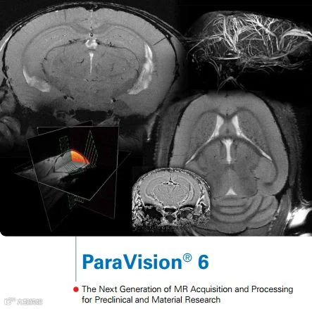

ParaVision® sets a new standard in preclinical imaging software. Its unique features enable easy and accelerated workflow for routine users and experts. With its well structured library of in vivo optimized protocols, its sophisticated image processing, visualization and data analysis features, ParaVision 6 ensures maximum productivity enhancing biomedical and pharmaceutical research.

The ParaVision 6 workflow is designed for optimal operation of MRI systems and is powered by the new electronic AVANCE III HD MRI platform. This now enables automatic hardware recognition for fast and easy setup, integrated dynamic realtime optimization and adjustment of experimental parameters during scanning, dynamic shimming and fast 64 bit multi-core image reconstruction
for high resolution 3D imaging results.
All application packages benefit from ParaVision’s new fully searchable, easy to use data base, network data management, and new archiving capabilities enabling the connection of multiple ParaVision workstations and DICOM server archiving. ParaVision 6 also supports and facilitates data exchange with other imaging modalities in your lab.
ParaVision 6 highlights
-
New and accelerated, intuitive workflow
-
Ready-to-use protocols and scan programs
-
Active method conflict handling
-
Automatic hardware recognition
-
Real-time optimization during scanning
-
On-the-fly RF-pulse calculation
-
In-line navigator-based image reconstruction
-
Fast 64 bit multi core image reconstruction for high resolution 3D
-
Fully searchable data base, network data management

Optimized Workflow
The ParaVision 6 user interface of dynamic context menus, cards and icons offers instant access to the most commonly used imaging protocols and processing tools.
Unmatched portfolio of application packages
ParaVision 6 provides various dedicated, optional preclinical imaging packages for a wide range of animal MR imaging and spectroscopy applications.
All benefit from the new accelerated workflow incorporating ready-to-use protocols and scan programs combined with interactive method and parameter for conflict handling.
ParaVision application packages include:
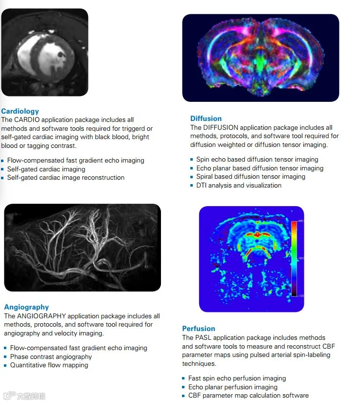
Cardiology
Diffusion
Angiography
Perfusion
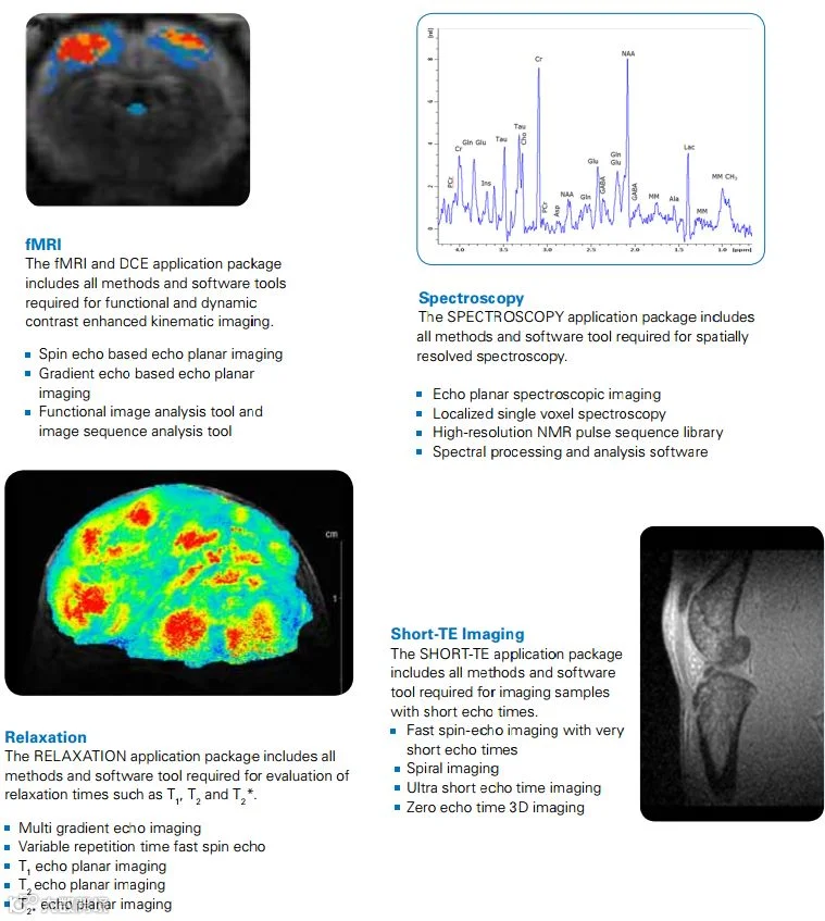
Spectroscopy
fMRI and DCE
Relaxation
Short TE Imaging
Method development framework
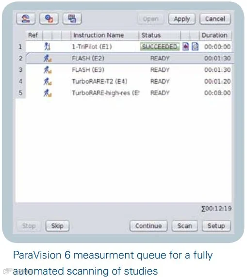
Automation
For maximized throughput ParaVision 6 provides fully automated acquisition and processing, including scan queueing. This productivity tool excels whenever high animal throughput accrues, or when your MRI system is accessed by many users.

3D Visualization and Analysis
3D data visualization and analysis tools extend basic viewing functionality:
3D interactive viewing
Maximum intensity projection (MIP)
Multi-planar secondary image reconstruction (MPR)
Segmentation
Surface rendering
Movie generation
Parallel Imaging and Reconstruction
Parallel imaging and reconstruction techniques including:
Accelerated parallel image acquisition
GRAPPA based image reconstruction
Parallel Transmit
Acquisition functionality for parallel transmit imaging techniques including:
Support of multi-channel parallel transmit RFpulses in all methods
Optimized phase calibration for homogeneous
excitation with multi-element RF transmitter coils
Parallel transmit template methods and protocols
3D Evaluation
Tools includes interactive magnification in real time, quantitative ROI analysis, geometric measurements, digital filtering, movie display of image sequences, and 3D secondary reconstruction of orthogonal planes.
3D imaging with 3D image processing
3D data evaluation and visualization e.g. multi planar reconstruction
MR angiography
Image algebra and image sequence analysis
Method Development Framework
A transparent method-programming framework including powerful tools, access to method toolboxes and method source code enable easy implementation of image acquisition and dedicated reconstruction techniques with only basic programming knowledge.
DTI Evaluation
3D region growing for signal masking
2D & 3D visualization of composite images
DTI-EPI supporting k-space trajectory based reconstruction
Calculation module to derive scalar and directional information
Visualization of direction-encoded color-maps in 2D and 3D together with morphological reference images
Transparency modulation by fractional anisotropy images
Arbitrary composition schemes and color coding capabilities
Network Data Management and Archiving
The database browser functionality enables network data transfer between multiple ParaVision workstations, facilitates data exchange with other imaging modalities in your lab and provides archiving on DVD and to DICOM server.
Multimodal Data Import
Import DICOM data to ParaVision from the following modalities:
Magnetic Resonance Imaging (MRI)
Positron – Emission Tomography (PET)
Computer Assisted Tomography (CT)
Magnetic Particle Imaging (MPI)
Single Positron Emission Computed
Tomography (SPECT)
Project Planning
Off-line registration and preparation from a remote processing workplace, including:
Subjects
Sessions, studies, scans and projects
Protocols
Scan programs

Pioneering Preclinical Imaging Solutions

点击“阅读原文”至布鲁克官网观看回放