
点击公共号即可订阅
Bone imaging and analysis with microCT: Exploring the possibilities
Seminar overview
Micro-computed tomography, or micro-CT, is being employed by a rapidly increasing number of laboratories for bone research. This webinar will give a better understanding of the methodology with its capabilities and limitations. The background of micro-CT bone analysis will be outlined, along with a introductory look at physical principles underlying the technique.
There is a brief examination of the methodology of two important types of micro-CT analysis - morphometric and density measurement. Finally, there will be a review of some selected applications of micro-CT in published bone research, including both ex-vivo and in-vivo analysis of bone.
Date
August 03, 2016
What will you discover
This webinar will cover the basics of MicroCT technology and give an overview of the possibilities of microCT in bone imaging and analysis.
Key learnings
Non-invasive 3D visualization and measurement with the micro-computed tomography (micro-CT)
How micro-CT can be applied to bone research
The capabilities of micro-CT methodology
-
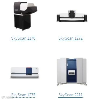
Ex-vivo and in-vivo analysis of bone research with the SkyScan systems
Who should attend
Any Lab Managers, Scientists, Researchers, Technicians, Students working in Academia, Hospitals or Institutions currently interested in the expanding field of microCT for preclinical Applications.
Research group leaders
Laboratory managers
Biomedical Scientists
microCT Specialist
Oncology Scientists
Bone Researchers
-
Cardiology Scientist
Presenters

Kjell Laperre, PhD - Applications Manager, Bruker microCT (biography)

点击“阅读原文”至布鲁克官网观看回放