

很长一段时间以来,科学家们一直依靠平板培养的2D细胞来研究细胞和疾病的机制。2D细胞模型对于细胞培养和处理当然简单且经济。
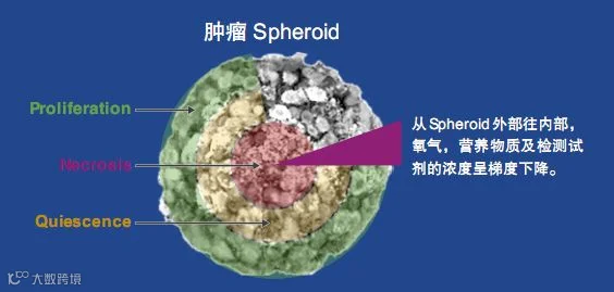
然而,我们可以看到在过去的十年里,3D细胞培养越来越受欢迎,因为它们在生理上更为相关,更能代表体内组织。仔细思考,我们体内没有一种细胞以独立于其他细胞或组织的形式进行单层生长。相反,大多数细胞自然存在于复杂的三维结构中,包括细胞外基质中的不同细胞类型。众多的细胞-细胞和细胞-基质相互作用都对它们的行为有着深刻的影响。此外,2D单分子膜可以均匀地获得营养和氧气,而肿瘤等细胞团则不是这样。3D肿瘤球体更能代表体内肿瘤,与外层相比,内部细胞获得营养和氧气的机会更少,形成自然梯度。
类器官、球状体和3D细胞模型研究在包括疾病建模和再生医学在内的许多应用中表现出了巨大的潜力。相对于2D模型,类器官和球状体等3D细胞模型使我们有机会在生理学相关背景下更好地理解生物学的复杂性。经过验证的实验方案和教育资源增强了我们对于培养和分析类器官和球状体的信心,引领3D模型取得成功。

3D 培养的细胞进行分析时面临的一个挑战是:找到适合你细胞类型的分析方法。大多数现有的基于细胞的分析最初是为 2D 单层或悬浮细胞设计,其设计并不考虑 3D 结构的大小或质量。在许多情况下,细胞外基质(ECM)层可以阻止试剂渗透到微球体 (Microspheroid) 中心,这就是为什么优化和验证检测特定的 3D 模型的细胞学分析系统变得至关重要。Promega 提供的商品化 3D 模型检测系统将为您解决这个问题,发光法的高灵敏度检测方案,快速检测 3D 模型的细胞健康,能量代谢等相关指标。
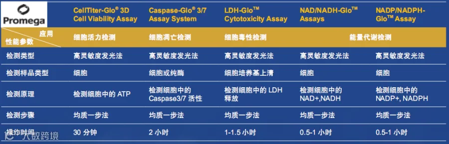
3D细胞活力检测 CellTiter-Glo® 3D Cell Viability Assay
3D细胞毒性检测 LDH-GloTM Cytotoxicity Assay
3D细胞能量代谢检测 NAD/NADH-GloTM Assay
3D细胞能量代谢检测 NADP/NADPH-GloTM Assay
为何要使用3D培养细胞模型
3D培养物更好地模拟组织样结构
能够表现出不同的细胞功能
可以共培养两种或多种不同的细胞类型
可模拟微环境条件,如缺氧和营养梯度
3D培养能够独立评估微环境的不同特征如何调节组织器官发生和疾病
更好地预测药物治疗的体内反应
3D培养是基础、转化和临床研究的自然整合点
3D细胞培养的应用
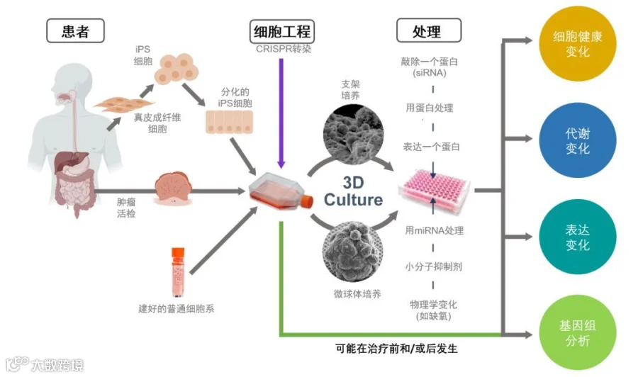
3D细胞模型越来越多地被用来了解疾病机制和药物研发治疗。这个过程可能涉及从患者身上提取组织细胞进行3D培养,如肿瘤类器官。3D培养物可以用来小分子药物筛选或者通过基因操纵来了解疾病的途径。与2D培养相比,3D细胞培养更准确地预测药物治疗的疗效或毒性。
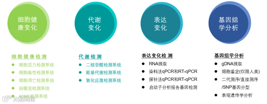
监测3D培养物的生物学变化
3D培养细胞学检测的挑战
使用3D培养的一个挑战是找到适合你细胞类型的分析方法。大多数现有的基于细胞的分析最初是为2D单层或悬浮细胞设计,可能并不考虑三维结构的大小或质量,这可能限制细胞裂解或试剂的化学成分设计。在许多情况下,细胞外基质(ECM)可以防止试剂渗透到球体的中心。这就是为什么为您的特定3D模型系统优化和验证基于细胞的分析是至关重要的。Promega为您提供用于3D培养细胞模型从细胞健康分析到基因组学分析的多种分析方法,助力您的研究。

3D培养检测设计与优化应考虑的因素
细胞裂解
成像的限制
非裂解性探针试剂的穿透性
细胞质量可能影响检测试剂的化学反应

CellTiter-Glo ® Luminescent Cell Viability Assay是基于ATP检测的快速细胞活力检测法。在国内外被广泛应用和公认的高灵敏度发光检测法,细胞活力检测的金标准。而CellTiter-Glo® 3D Cell Viability Assay基于经典CellTiter-Glo检测原理,专门为检测3D微组织球的细胞活力而优化。该检测提供的试剂可以高效的渗透至大的细胞球,相比常规细胞活力检测试剂,具有更强的裂解能力,从而为细胞活力的测定提供更加准确的结果。
已成功于多种方式培养的3D微组织的活力检测,包括:
细胞悬滴培养板(hanging-drop plate)
超低吸附细胞培养板
Matrigel™ 包被的细胞培养板
琼脂糖包被细胞板
甲基纤维素覆盖培养液
Alvetex® 三维细胞培养体系
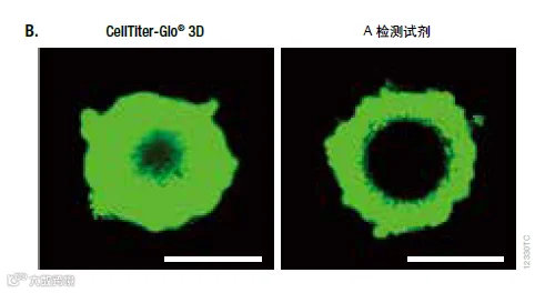
将不同数量的HCT116细胞(RPMI培养基+10% FBS) 接种在InSphero GravityPLUSTM96-孔悬滴培养板, 培养4天。
图A. 在微组织培养孔中,加入与培养基等体积的CellTiter-Glo® 3D或其他厂家的A检测试剂,震荡混匀5分钟后,室温孵育30分钟,应用GloMax®发光检测仪读数。
图B. 2X CellTox™ 绿色荧光染料( 结合于膜完整性受损的细胞的双链DNA) 提前预混在CellTiter-Glo® 3D 检测试剂(左)或A检测试剂(右)中,后共同加入细胞培养孔。震荡混匀5分钟后,室温孵育30分钟,应用激光共聚焦显微镜拍照。绿色荧光染色显示裂解的细胞。微组织直径为300μm。

1.3D tumor spheroid models for in vitro therapeutic screening: a systematic approach to enhance the biological relevance of data obtained, Scientific Reports, 2016
2.Patterning of tissue spheroids biofabricated from human fibroblasts on the surface of electrospun polyurethane matrix using 3D bioprinter, International Journal of Bioprinting, 2016
3.Inhibition of monocarboxylate transporter 1 suppresses the proliferation of glioblastoma stem cells, The Journal of Physiological Sciences, 2016
4.Influence of Matrices on 3D-Cultured Prostate Cancer Cells' Drug Response and Expression of Drug-Action Associated Proteins, Plos one, 2016
5.cRGD peptide installation on cisplatin-loaded nanomedicines enhances efficacy against locally advanced head and neck squamous cell carcinoma bearing cancer stem-like cells, Journal of Controlled Release, 2017
6.Monitoring the effects of doxorubicin on 3D-spheroid tumor cells in real-time, Onco Targets Ther, 2016
7.Prolyl isomerase Pin1 promotes survival in EGFR-mutant lung adenocarcinoma cells with an epithelial–mesenchymal transition phenotype, Laboratory Investigation, 2016
8.Drug Discovery Goes Three-Dimensional: Goodbye to Flat High-Throughput Screening? ASSAY and Drug Development Technologies, 2015
9.A bioengineered three-dimensional cell culture platform integrated with microfluidics to address antimicrobial resistance in tuberculosis, mBio, 2017
10.Impact of adjustable cryogel properties on the performance of prostate cancer cells in 3D, SpringerPlus, 2016
11.Silk scaffolds connected with different naturally occurring biomaterials for prostate cancer cell cultivation in 3 D, Biopolymers, 2016
12.A High-Content Screening of Anticancer Compounds Suggests the Multiple Tyrosine Kinase Inhibitor Ponatinib for Repurposing in Neuroblastoma Therapy, AACR Journals, 2018
13.Overactive mTOR signaling leads to endometrial hyperplasia in aged women and mice, Molecular cancer therapeutics, 2017
14.Quantitative high-throughput phenotypic screening of pediatric cancer cell lines identifies multiple opportunities for drug repurposing, Oncotarget, 2018
15.Elevation of sensitivity to anticancer agents of human lung adenocarcinoma A549 cells by knockdown of claudin-2 expression in monolayer and spheroid culture models, Biophysica Acta, 2018
16.Miniring approach for high-throughput drug screenings in 3D tumor models, bioRxiv, 2017
17.A PDX/organoid biobank of advanced prostate cancers captures genomic and phenotypic heterogeneity for disease modeling and therapeutic screening, Clinical cancer research, 2018
18.C3aR and C5aR1 act as key regulators of human and mouse β-cell function, Cellular and Molecular Life Sciences, 2018
19.Establishment of patient-derived tumor spheroids for non-small cell lung cancer, PLOS one, 2018
20.Evaluation of anticancer agents using patient-derived tumor organoids characteristically similar to source tissues, Oncology Reports, 2018
21.DGC-specific RHOA mutations maintained cancer cell survival and promoted cell migration via ROCK inactivation, Oncotarget, 2018
22.A novel three-dimensional high-throughput screening approach identifies inducers of a mutant KRAS selective lethal phenotype, Oncogene, 2018
23.Cytotoxicity evaluation using cryopreserved primary human hepatocytes in various culture formats, Toxicology letters, 2016
24.Endogenous replication stress marks melanomas sensitive to CHK1 inhibitors in vivo, Clinical cancer research , 2018
25.Toxicology studies of primycin-sulphate using a three-dimensional (3D) in vitro human liver aggregate model, Toxicology letters, 2017