

抗原分子表面通常分布着多个抗原决定簇(epitopes),两个分别针对同一抗原分子上不同抗原决定簇的抗体,能够同时结合到该抗原上,形成配对抗体。
然而,实际情况远比理论复杂,即使两个抗体针对的是同一抗原的不同抗原决定簇,也并不意味着这两个抗体一定可以同时与该抗原结合,成为抗体对。当一个抗体与抗原结合后,可能会引发抗原表面构象的变化,从而阻碍其他抗体的结合[3]。空间位阻效应也可能成为障碍,尤其是当两个抗原决定簇在三维空间上距离较近时,可能无法同时容纳两个抗体分子进行有效结合[4]。
因此,成功制备出能够同时结合于同一抗原分子的配对抗体,并非易事。这不仅要求抗体设计上的精准与巧妙,在抗体制备后,还要进行一系列严格的筛选和验证。
常规抗体缓冲体系中往往含有牛血清白蛋白(BSA)、叠氮化钠(NaN3)及甘油。BSA会与一抗竞争结合偶联物,影响抗体标记效率。NaN3则会抑制辣根过氧化物酶(HRP)的活性,同时具有细胞毒性,会限制抗体在细胞培养中的使用。抗体缓冲体系中的甘油浓度过高会影响分子扩散,导致染料分子和抗体分子不能充分结合。Proteintech抗体对去除了这些组分,仅含有PBS。
2. ABCE™重组抗体平台开发,高通量,高质量
Proteintech抗体对绝大多数采用重组兔单抗技术,基于ABCE™单个B细胞抗体发现技术平台(点击蓝字了解详情)研发的重组兔单抗具有特异性高,批间一致性高等优点。这是Proteintech科学家们能筛选出好的抗体对的基础。
3. 天然样本/亲和力/CBA/ELISA验证四重验证,确保高特异性
Proteintech抗体对均通过CBA技术进行筛选并验证,大部分热门靶点同时经过ELISA检测进行双重验证,其他靶点也正在逐步验证中。正在逐步对所有样本做天然样本验证。
4. 数量庞大的抗体对库:灵活应对免疫检测挑战
Proteintech目前有7000+种抗体对,覆盖自身免疫疾病、心血管标志物、糖尿病、消化和代谢类、炎症标志物、肿瘤标志物等常见疾病关键靶点。
部分数据展示
以TREM-1/CD354重组抗体对(货号:MP00988-1)为例
● 天然样本检测数据

◀4份健康人唾液样本检测图。检测样品的TREM-1/CD354浓度为65.0 pg/mL,范围为4.0-111.2 pg/mL
▶8份健康人血清样本检测图。检测样品的TREM-1/CD354浓度为230.6 pg/mL,范围为113.8-438.7pg/mL

● 亲和力参数数据

◀TREM-1/CD354重组捕获抗体(84071-3-PBS)的亲和力和动力学表征,亲和常数为1.48 nM
▶TREM-1/CD354重组检测抗体(84071-2-PBS)的亲和力和动力学表征,亲和常数为 6.13 nM

● CBA及双抗夹心ELISA测定数据

◀MP00988-1的CBA标准曲线,检测范围:0.156-20 ng/mL
▶MP00988-1的双抗夹心ELISA标准曲线,检测范围:3.91-250 pg/mL

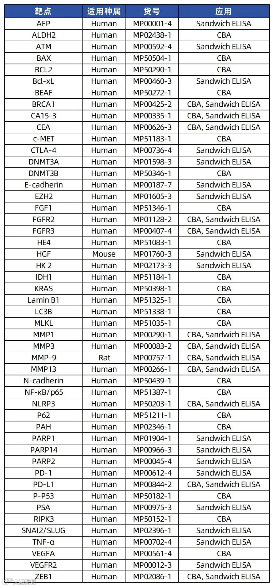
热门靶点解析
小P这一期带来肿瘤生物标志物、炎症反应生物标志物、急性冠脉综合征标志物等领域热门靶点解析。
肿瘤生物标志物是肿瘤发生和发展过程中由肿瘤或机体对肿瘤的反应产生的物质,使用抗体对检测肿瘤生物标志物在癌症的筛查和早期诊断、预后预测、复发检测和治疗效果监测方面具有重要价值。根据检测样本类型和来源,我们可以将肿瘤生物标志物分成三类:来自血液、组织和其他生物液体(如尿液和唾液)的肿瘤生物标志物。
1.1 血液中的肿瘤标志物
AFP(抗体对货号:MP00001-4):AFP被认为是胎儿时期血清白蛋白的类似物,是胎儿循环的主要蛋白质。健康成人血清中AFP浓度低于10 μg/ L[1]。AFP是目前应用最广泛的HCC肿瘤生物标志物,国际学术界建议将AFP的参考值限制在20 μg/L。HCC患者AFP急剧升高提示肿瘤复发或转移。手术后AFP >200 μg/L提示HCC未完全切除或转移[2-3]。
CEA(抗体对货号:MP00626-3):CEA属于细胞表面的糖蛋白家族,是一种广谱肿瘤生物标志物,CEA在70%的结直肠癌、55%的胰腺癌、50%的胃癌、45%的肺癌、40%的乳腺癌、40%的尿道癌和25%的卵巢癌患者中升高。血清CEA水平与肿瘤负荷成正比。CEA在肿瘤患者中被用于辅助诊断、判断预后、监测复发和评价治疗效果[4-7]。
PSA(抗体对货号:MP00975-3):PSA是前列腺上皮细胞特异性分泌的丝氨酸蛋白激酶释放酶,由kallikrein 3编码。对于前列腺癌的早期诊断,血清PSA阳性临界值大于10 ng/mL。然而,PSA在前列腺癌诊断中的特异性较差,一些非癌性病变,如炎症、创伤或良性前列腺增生也可能升高PSA水平,从而导致高假阳性率。综上所述,PSA水平在前列腺癌的诊断和预测中是一个很有前景的生物标志物[8-10]。
CA15-3(抗体对货号:MP00335-1):糖类抗原15-3 (CA15-3,又称粘蛋白1)是一种由MUC1基因衍生的大型跨膜糖蛋白。并正常血清CA15-3水平的参考值小于28 U/mL。在乳腺癌中,CA15-3联合CEA是最常用的乳腺癌诊断方法。CA15-3也是评价乳腺癌术后恢复、复发、转移的重要指标[11-12]。
HE4(抗体对货号:MP51083-1):人附睾蛋白4 (HE4)是一种乳香酸蛋白,广泛表达于气管、唾液腺、肺组织等,在卵巢癌、子宫内膜癌、肺癌中均有高表达。目前,血清HE4主要用于卵巢癌的诊断和复发监测,敏感性为67%[13-15]。
1.2 组织中的肿瘤标志物
细胞增殖相关:癌细胞能够通过多种途径获得维持增殖的能力,包括RAS、磷PI3K-AKT-mTOR、RAF-MEK-ERK通路。
KRAS(抗体对货号:MP50398-1):KRAS是迄今为止三种RAS基因中最常扩增和突变的RAS亚型,占所有RAS突变的85%。KRAS突变存在于88%的胰腺癌、50%的CRC和32%的肺癌中[16-17]。
BEAF(抗体对货号:MP50272-1):BRAF突变在癌症中被广泛研究,在ER阴性或孕激素受体阴性的乳腺癌患者中,BRAF-MEK-ERK通路的高活性与较差的生存率相关[18-19]。
PTEN(抗体对货号:MP00328-6):PTEN是一种磷酸肌苷3-磷酸酶,负向调控PI3K-AKT-mTOR通路,作为各种癌症的预后和预测性生物标志物,包括前列腺癌、RCC、PDAC、CRC、乳腺癌、子宫内膜癌、脑癌、皮肤癌和血液恶性肿瘤[20]。
抑癌基因相关:肿瘤细胞避开了强大的程序来逃避生长限制和阻断,包括Rb、TP53等信号通路。
血管生成相关:肿瘤的发生和发展过程中,新的血管系统可以运输营养和氧气,并排泄代谢废物。血管生成因子如VEGF等诱导内皮细胞的增殖和分化。
VEGF与和VEGFR2(抗体对货号:MP00012-3):VEGF和VEGFR-2是预测预后和抗血管生成药物疗效的重要生物标志物,VEGF水平与肿瘤预后呈负相关。此外,VEGFR抑制剂sorafenib对高水平VEGF的晚期透明细胞肾细胞癌患者显示出更好的治疗效果[21-23]。
FGF与FGFR:FGF1(抗体对货号:MP51346-1)是卵巢癌的预后生物标志物,在高级别浆液性卵巢癌中,FGF1扩增诱导的血管生成增加导致患者OS降低。在超过30%的膀胱癌中观察到FGFR3(抗体对货号:MP00407-4)突变,在12%的子宫内膜癌中发现了FGFR2(抗体对货号:MP01128-2)突变。大约10%的胃癌表现出FGFR2扩增,这与胃癌患者预后不良有关[24-27]。
抵抗细胞死亡相关:抵抗细胞死亡是肿瘤的一个重要标志。在高度侵袭性和治疗耐药的肿瘤细胞中,包括细胞凋亡相关如BCL-2、BAX等,自噬相关如LC3B、p62等,细胞坏死相关包括RIPK3、MLKL等发生改变。
BCL-2(抗体对货号:MP50290-1)/BCL-XL(抗体对货号:MP00460-3):BCL-2是第一个被发现的细胞凋亡调节因子,是TNBC患者的预后生物标志物。BCL-XL是BCL-2蛋白家族成员,可作为预测结直肠癌患者预后的独立生物标志物[28-29]。
BAX(抗体对货号:MP50504-1):在包括胃癌、食管癌和CRC在内的多种癌症中,BAX基因作为一种潜在的预后和预测性生物标志物。BAX表达降低是卵巢癌细胞对顺铂耐药、CRC细胞对5-FU耐药、肺癌细胞对唑来膦酸盐耐药的主要因素[30-33]。
LC3B(抗体对货号:MP51338-1):经典的自噬标志物,LC3B的高表达与胃癌、CRC、TNBC、黑色素瘤、星形细胞瘤、食管癌、等多种肿瘤的侵袭性进展及不良预后密切相关。此外,LC3B与HCC的血管侵袭和淋巴结转移密切相关,是HCC的潜在治疗靶点[34-40]。
p62(抗体对货号:MP51211-1):p62高表达与癌症的侵袭性和不良预后相关,包括子宫内膜癌、OSCC、上皮性卵巢癌和NSCLC。此外,p62高表达还与乳腺癌患者的高级别、远处转移和5年生存率降低相关,尤其是TNBC癌症患者[41-44]。
RIPK3(抗体对货号:MP50152-1):RIPK3需要根据具体情况进行分析,在多种肿瘤细胞中下调,包括乳腺癌、黑色素瘤、肺癌和CRC,但在其他几种肿瘤中表达升高,如浆液性卵巢癌、胰腺癌、结肠炎相关癌和结肠癌[45-51]。
MLKL(抗体对货号:MP51035-1):MLKL的低表达水平与胃癌、卵巢癌、宫颈癌、结肠癌以及胰腺癌的低OS显著相关。在接受辅助化疗的PADC切除患者中,MLKL低表达水平与RFS降低有关。因此,MLKL已成为早期PADC切除患者的预后生物标志物[52-56]。
侵袭和转移相关:活化侵袭和转移已被认为是肿瘤的标志之一,也是实体瘤患者死亡的主要原因,这个过程依赖于E-cadherin、SLUG等基因。
E-cadherin(抗体对货号:MP00187-7):E-cadherin的缺失或下调可促进肿瘤侵袭、浸润性生长和去分化。E-cadherin可作为多种肿瘤转移的预后生物标志物[57]。
SLUG(抗体对货号:MP02396-1):Snail家族的一员,对EMT有显著的影响。SLUG表达是CRC661和食管SCC患者生存不良的独立预后生物标志物[58-59]。
ZEB1(抗体对货号:MP02086-1):ZEB1的异常表达与多种肿瘤的进展和转移有关,包括子宫癌、骨肉瘤、肺癌、肝癌和胃癌[60]。
HGF(抗体对货号:MP01760-3)/c-MET(抗体对货号:MP51183-1):c-MET是原癌基因,HGF是c-MET配体,c-MET的转录失调、降解不足和HGF的异常产生与肿瘤的进展密切相关。c-MET表达与结直肠癌肝转移的肿瘤分期呈正相关。除胃肠道肿瘤外,乳头状肾癌、卵巢癌、SCLC、 HNSCC和儿童HCC中也发现了c-MET突变。HGF水平升高存在于头颈癌、宫颈癌、HCC和肺癌等多种癌症中,且与预后不良有关。HGF/c-MET可作为多种血液肿瘤的预后生物标志物,如b细胞淋巴瘤、T细胞和自然杀伤细胞淋巴瘤以及霍奇金淋巴瘤[61-73]。
N-cadherin(抗体对货号:MP50439-1):N-cadherin在多种肿瘤中高表达,包括黑色素瘤、神经母细胞瘤、乳腺癌、尿路上皮癌、卵巢癌、多发性骨髓瘤等。此外,N-cadherin在血液学恶性肿瘤(如白血病和多发性骨髓瘤)中表现出重要作用,并与多发性骨髓瘤的不良预后密切相[74-80]。
MMPs:在不同肿瘤中均观察到MMPs的上调。MMP9(抗体对货号:MP00757-1)在癌细胞的侵袭和转移中起着至关重要的作用,并已被证明是不同癌症的关键生物标志物,包括NSCLC、宫颈癌、胃癌、卵巢癌、乳腺癌、骨肉瘤和胰腺癌。MMP1(抗体对货号:MP00290-1)、MMP2(抗体对货号:MP51054-1)和MMP16的表达与葡萄膜黑色素瘤患者的OS和DFS呈正相关[81-88]。
突变相关:基因组不稳定和突变是癌症的重要标志,这个过程需要PARP等基因参与。
PARP:PARP在许多类型的癌症中起重要作用,包括卵巢癌、乳腺癌、胰腺癌和前列腺癌。PARP抑制剂可以阻断SSB修复途径,并在同源重组(HR)缺陷导致的DSB修复受损的癌症中引发致死性[89-91]。
BRCA1(抗体对货号:MP00425-2):在消化道癌症中,细胞质BRCA1和BRCA2的高表达与有利的OS显著相关,而BRCA1核表达通常预示不良预后。此外,BRCA1/2突变与多种癌症的进展密切相关,包括乳腺癌、卵巢癌、前列腺癌和胰腺癌[92]。
ATM(抗体对货号:MP00592-4):ATM在多种肿瘤中经常发生突变或失活。携带ATM突变的子宫内膜癌患者表现出更高的肿瘤突变可能和更高的免疫检查点表达水平。此外,ATM突变与转移性CRC患者的生存期延长独立相关[93-94]。
肿瘤促进炎症相关:炎症通过控制癌症的发展、血管生成、恶性转化、侵袭和迁移、免疫监视和对治疗的反应,在各种人类癌症中起重要的作用。炎症相关调节因子,包括TNF-α(抗体对货号:MP00702-4)、NF-κB(抗体对货号:MP51387-1)和NLRP3(抗体对货号:MP50203-1),是潜在的肿瘤预后生物标志物。
代谢重编程相关:肿瘤细胞也会通过将能量代谢主要限制为糖酵解来重编程糖代谢,从而重编程能量产生,这个过程依赖于HK2、IDH1/2等基因参与。
HK2(抗体对货号:MP02173-3):HK2过表达与实体肿瘤中更差的OS和PFS显着相关。在HCC、胃癌和CRC患者中观察到HK2对OS的负面影响。HK2表达与晚期和高级别卵巢癌相关,HK2下调抑制肿瘤发生[95-97]。
IDH1/2(抗体对货号:MP51184-1):IDH1和IDH2突变发生在多种血液学和实体肿瘤中,包括胶质瘤、AML、肝内胆管癌、软骨肉瘤、甲状腺癌和血管免疫母细胞T细胞淋巴瘤[98-100]。
免疫逃逸相关:癌细胞通过破坏免疫系统产生免疫逃逸,最终促进肿瘤的进展、扩散和转移,涉及PD-1/PD-L1、CATL-4等基因。
PD-1(抗体对货号:MP00612-4)/PD-L1(抗体对货号:MP00844-2):激活的PD-1/PD-L1信号负调控外周组织中T细胞介导的免疫反应,从而限制效应T细胞反应并保护组织免受损伤。阻断PD-1/PD-L1饥饿和的免疫检查点抑制剂有效地延长了各种癌症患者的生存期。
CTLA-4(抗体对货号:MP00736-4):CTLA-4促进肿瘤逃避宿主免疫监视,并参与多种癌症的免疫失调,包括肺癌、宫颈癌、乳腺癌、皮肤癌、胃癌、CRC、B细胞CLL和非霍奇金淋巴瘤[101]。
去分化相关:分化和去分化在许多肿瘤的发育过程中也是必不可少的。分化相关基因,如ALDH2(抗体对货号:MP02438-1)、PAH(抗体对货号:MP02346-1)等,已被鉴定为预测多种癌症的生存和不良预后[102]。
甲基化相关:DNA甲基化的生物标志物已被誉为癌症生物标志物研究中的一个重要事件,DNA甲基化主要由三种DNA甲基转移酶DNMT1、DNMT3A(抗体对货号:MP01598-3)和DNMT3B(抗体对货号:MP50346-1)催化。DNMTs在多种癌症中过表达,包括AML、CML、胶质瘤、乳腺癌、胃癌、CRC、HCC、胰腺癌、前列腺癌和肺癌[103-104]。
组蛋白修饰相关:组蛋白修饰总体水平的变化可以预测各种癌症的临床结果,组蛋白修饰是一个动态过程,受写入器如组蛋白乙酰转移酶、组蛋白甲基转移酶、读取器如含溴结构域的蛋白质,以及擦除器如组蛋白去乙酰化酶和赖氨酸去甲基化酶控制。
EZH2(抗体对货号:MP01605-3):EZH2负责H3K27的甲基化。EZH2过表达与前列腺癌和乳腺癌的不良预后密切相关。髓系恶性肿瘤和T细胞急性淋巴细胞白血病中EZH2基因的功能缺失突变也会导致预后不良[105-106]。
细胞衰老相关:细胞衰老相关基因是肿瘤细胞的重要生物标志物。衰老相关分泌表型(SASP)激活,SASP能够以旁分泌的方式将信号分子传递给邻近的活癌细胞以及TME中的其他细胞。NF-kB调控的SASP因子具有肿瘤抑制和免疫监视作用,而信号换能器和转录激活因子3 (STAT3)调控的SASP因子具有肿瘤促进和免疫抑制作用[107]。衰老细胞中发现层Lamin B1(抗体对货号:MP51325-1)的表达减少,肿瘤组织中广泛观察到Lamin B1上调。Lamin B1在HCC中过表达并促进细胞增殖和转移,Lamin B1表达升高表明HCC预后和免疫治疗反应较差。此外,Lamin B1已被提出作为ccRCC877和肺腺癌的预后衰老生物标志物[108-110]。
1.3 其他生物液体的肿瘤生物标志物
液体活检已成为癌症诊断、实时监测和通过微创检测生物液体(如血液、唾液、尿液、胸膜液和腹水)进行预后的关键策略。液体活检肿瘤诊断生物标志物,包括循环肿瘤DNA、循环肿瘤细胞和外泌体,都是肿瘤诊断和治疗的有效监测工具。
(↓上下滑动以查看全部↓)
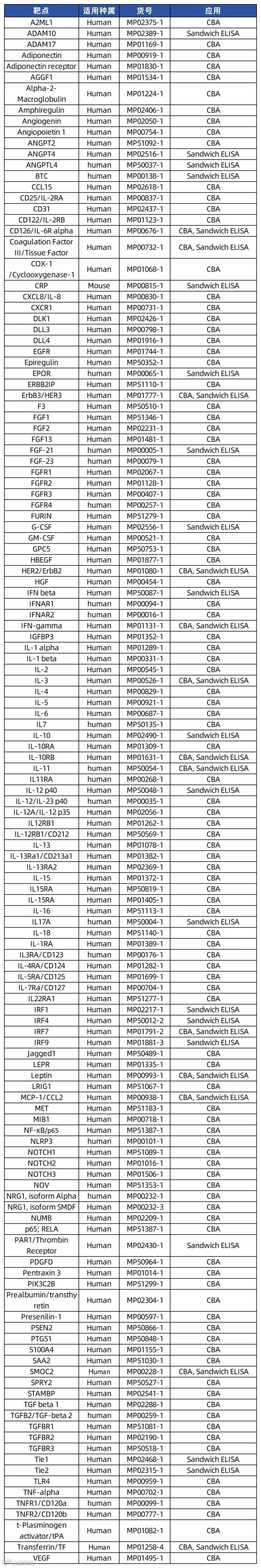
炎症的生物标志物通常是血清、血浆或血液来源的蛋白质或酶,它们具有独立的诊断和预后价值,代表了疾病的潜在状态,包括促炎细胞因子和急性时相蛋白APPs。除此之外,还有一些次要的炎症标志物,如转录因子NF-κB等。
细胞因子:细胞因子(cytokine,CK)是免疫原、丝裂原或其他刺激剂诱导多种细胞产生的低分子量可溶性蛋白质,具有调节固有免疫和适应性免疫等多种功能。细胞因子可被分为白细胞介素、干扰素、肿瘤坏死因子超家族等。
IL-6(抗体对货号:MP00687-1):IL-6是一种白细胞介素,作为促炎和抗炎细胞因子。IL-6蛋白是包括T细胞和巨噬细胞在内的多种细胞类型分泌的磷酸化和可变糖基化分子。IL-6在B细胞向IgG分泌细胞的最终分化过程中起重要作用,参与淋巴细胞和单核细胞的分化。
IL-8(抗体对货号:MP00830-1):白细胞介素8 (IL-8),也称为CXCL8,是CXC趋化因子家族的一员。这种趋化因子由多种细胞类型分泌,包括单核/巨噬细胞、T细胞、中性粒细胞、成纤维细胞、内皮细胞和各种肿瘤细胞系,以响应炎症刺激。该基因被认为在毛细支气管炎的发病机制中起作用,毛细支气管炎是一种由病毒感染引起的常见呼吸道疾病。IL-8升高与红斑狼疮、肝细胞癌、牙周炎有关[111-113]。
IFN-γ(抗体对货号:MP50092-1):IFN-γ是一种可溶性细胞因子,是II型IFN的唯一成员。它由Th1细胞、细胞毒性T细胞和NK细胞分泌。IFN-γ是巨噬细胞的有效激活剂,在病原体清除中起着至关重要的作用。异常的IFN-γ表达与许多自身炎症和自身免疫性疾病有关。它已在许多研究中被确定为胸膜结核(TB)的生物标志物。
TNF-α(抗体对货号:MP00702-4):TNF-α是一种多功能促炎细胞因子,主要由活化的巨噬细胞产生。它可以结合并通过其受体TNFRSF1A/TNFR1和TNFRSF1B/TNFBR发挥作用。这种细胞因子参与多种生物过程的调控,包括细胞增殖、分化、凋亡、脂质代谢和凝血。这种细胞因子与多种疾病有关,包括自身免疫性疾病、胰岛素抵抗和癌症。
急性时相反应蛋白:急性时相反应蛋白是一组在组织损伤、急性感染或炎症反应过程中浓度显著变化的血浆蛋白质,其合成主要由肝细胞在糖皮质激素及细胞因子(如IL-6、TNF-α)调控下完成 。正向成员包括C反应蛋白(CRP)、血清淀粉样蛋白A(SAA)等,负向成员则有前白蛋白、转铁蛋白等。
CRP(抗体对货号:MP00815-1):CRP由肝脏在炎症期间对白细胞介素-6 (IL-6)的反应中合成。CRP显示出与宿主防御相关的几种功能:它通过钙依赖性结合磷胆碱促进凝集、细菌荚膜肿胀、吞噬和补体固定。PD、冠心病和中风患者血液中水平升高[114-115]。
SAA:SAA蛋白是一组载脂蛋白,主要存在于血浆高密度脂蛋白(HDL)部分,主要由肝细胞产生,受炎症因子的调节。临床上常将SAA水平测定用于感染性疾病的诊断和病情监测,如儿童肺炎、腮腺炎等,再结合白细胞计数、C-反应蛋白等其他炎症指标,综合分析患者可能感染的病原体,非感染性疾病则常单独化验SAA,起辅助诊断和病情分析之用,如冠心病、类风湿关节炎等。
Haptoglobin(抗体对货号:MP00779-1):触珠蛋白是一种由肝脏合成的急性期反应蛋白,主要功能是结合游离血红蛋白,形成复合物后被单核-吞噬细胞系统清除,从而防止铁流失并减少血红蛋白对肾脏的损伤。炎症性疾病、吸烟者、肾病综合征和类风湿性关节炎中,触珠蛋白也会增加[116-117]。
Prealbumin(抗体对货号:MP02304-1):前白蛋白(Prealbumin,PAB),又称转甲状腺素蛋白(transthyretin,TTR),测定其在血浆中的浓度对于了解蛋白质的营养不良、肝功能不全、比之白蛋白和转铁蛋白具有更高的敏感性。前白蛋白还是一种急性负时相反应蛋白,在机体应激反应、坏死物质清除、组织修补等生理过程中扮演着重要角色,并且在心内科疾病的诊断、危险分层、预后评估等方面具有重要作用。
Transferrin:是血浆中主要的含铁蛋白质,负责运载由消化管吸收的铁和由红细胞降解释放的铁。以TRF-Fe3+的复合物形式进入骨髓中,供成熟红细胞的生成。TRF在急性时相反应中往往降低,在炎症、恶性病变时常随着白蛋白、前白蛋白同时下降。
转录因子:炎症信号激活由NF-κB和干扰素调节因子 (IRF) 家族转录因子介导的转录反应,从而产生促炎细胞因子、趋化因子和干扰素。
NF-κB(抗体对货号:MP01258-4):转录因子NF-κB普遍存在于几乎所有动物的各类细胞中,它参与调控一大类基因组的表达,包含免疫学有关受体、黏附分子如细胞间黏附分子、 血管细胞黏附分子和E选择素;炎症因子,粒细胞-巨噬细胞集落刺激因子以及急性期蛋白如血清淀粉样物质A等,因而促进人类的免疫学反应,在多种病理生理过程中扮演着至关重要的作用。
IRFs:干扰素调节因子(Interferon regulatory factors,IRFs)是一类重要转录因子家族,最初因调控I型干扰素(IFN-I)及其响应基因而被发现。IRFs通过协调抗病毒反应、促炎信号及细胞发育分化,在免疫稳态中发挥核心作用,同时兼具抑癌与促癌双重功能。该家族成员广泛参与感染性疾病、自身免疫病、代谢紊乱及肿瘤等多种病理过程。
(↓上下滑动以查看全部↓)
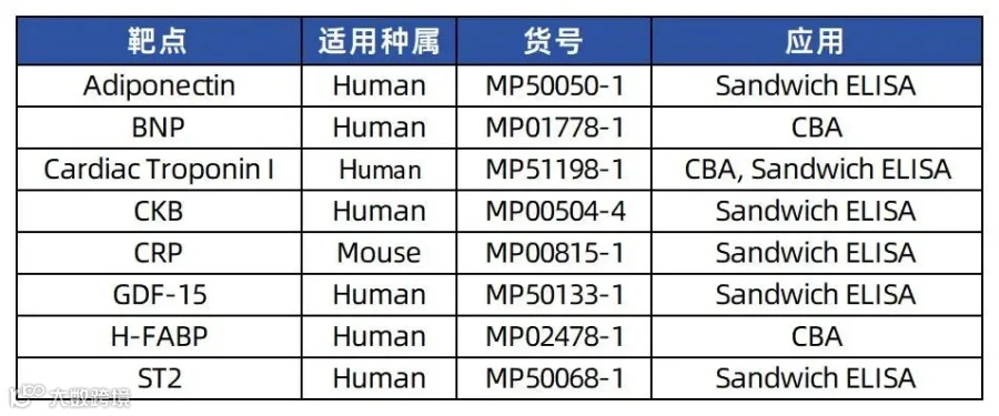
急性冠脉综合征(ACS)是一组由急性心肌缺血引起的临床综合征,包括不稳定型心绞痛、非ST段抬高型心肌梗死和ST段抬高型心肌梗死。心脏生物标志物是当心肌受损或受压时释放到血液中的内源性物质。 这些生物标志物的测量用于帮助诊断、评估风险和管理急性冠脉综合征。
H-FABP(抗体对货号:MP02478-1):心脏型脂肪酸结合蛋白(H-FABP)参与心肌细胞的脂肪酸代谢。H-FABP在症状出现后4小时内就诊的患者中检测AMI的敏感性为60%,在症状出现后4-12 h检测急性心肌梗死AMI的灵敏度为86.96%[118]。H-FABP在检测 AMI 方面的特异性较低,H-FABP不适合作为诊断AMI的独立检测,但作为特定患者群体的辅助检测可能具有一定的价值[119-120]。
CK-MB:肌酸激酶同工酶MB是由脑型亚单位(B)和肌型亚单位(M)组成的二聚体,主要存在于心肌组织中。心肌损伤后4小时在血清中检测到CK-MB,24小时达到峰值,48至72小时内恢复正常。 CK-MB2与CK-MB1的比率≥ 1.5,CK-MB相对指数(CK-MB/总CKx100)≥2.5 提高了心脏组织特异性并表明急性心肌梗死。由于 CK-MB 水平通常在心肌缺血后 48 至 72 小时内恢复正常(与肌钙蛋白相反,肌钙蛋白可以持续数天),监测CK-MB有助于确定水平在最初下降后是否再次升高[121-122]。
Cardiac troponin(抗体对货号:MP51198-1):心肌肌钙蛋白试验是评估疑似 AMI 患者的一线检查。肌钙蛋白是一种存在于心脏和骨骼肌中的蛋白质,在肌肉收缩中发挥作用。肌钙蛋白由 3 个亚基组成——肌钙蛋白 C、肌钙蛋白 I和肌钙蛋白 T。心脏中的肌钙蛋白 I 和肌钙蛋白 T 在结构上与骨骼肌中发现的肌钙蛋白 T 不同,使其成为心肌细胞损伤的特异性和敏感生物标志物。
CRP(抗体对货号:MP00815-1):CRP与血管功能障碍和动脉粥样硬化的进展有关,并已被证明可以预测未来的心血管事件,包括首次急性心肌梗死 (AMI)、中风和外周动脉疾病的发展[123]。
BNP(抗体对货号:MP01778-1):BNP基因表达的主要部位是心脏,并被翻译成亲本肽preproBNP。这种肽在细胞内质网的管腔内转运,在那里它被切割成proBNP和BNP信号肽。proBNP被分解为氨基末端产物NT-proBNP和生理活性BNP。BNP具有组成性释放,具有背景稳态和心脏保护作用。冠状动脉疾病越严重、缺血范围越大、左心室功能障碍(收缩和舒张)、局部壁运动异常和心力衰竭/心源性休克的患者BNP/NT-proBNP水平越高。BNP和NT-proBNP是AMI后不良事件(死亡、心力衰竭和复发性心脏缺血)的优秀标志物[124-131]。
GDF-15(抗体对货号:MP50133-1):生长分化因子-15 ( GDF-15)是转化生长因子-b细胞因子超家族的应激反应成员,参与调节各种器官发育、分化和组织修复所需的炎症和凋亡途径。动物模型显示,GDF-15在心肌缺血再灌注损伤、压力过载和心力衰竭时被诱导。GDF-15已被证明可以独立预测死亡率或死亡和非致命性AMI的组合,并对风险进行分级[132-134]。
Adiponectin(抗体对货号:MP50050-1):脂联素是一种脂肪细胞因子,是成熟脂肪细胞分泌的调节因子。它属于胶原超家族,与胶原、补体因子和TNF-α具有同源性。脂联素具有胰岛素增敏、抗炎、脂质代谢、抗动脉粥样硬化和抗血管生成作用。脂联素已被证明在内皮屏障受损后积聚在血管内皮下空间,在那里它抑制单核细胞粘附内皮细胞,抑制血管平滑肌的迁移和增殖(即它直接抗动脉粥样硬化)。脂联素水平在女性、肥胖者、2型糖尿病/胰岛素抵抗者、伴有高甘油三酯水平和低高密度脂蛋白水平的脂异常血症患者中较低,从而导致血管病变[135-138]。
ST2(抗体对货号:MP50068-1):ST2是一种具有白介素IL-1受体家族结构序列的肽,有2种同型,一种是膜结合形式ST2L,另一种是截断的可溶性形式。ST2L与IL33的结合也被证明在心肌过载的情况下被诱导,如心肌梗死和急性心力衰竭以及其他原因的心肌拉伸(左心室压力和容量过载)急性冠状动脉综合征患者(以及肺部疾病、急性心力衰竭、伴有和不伴有失代偿性急性心力衰竭和慢性心力衰竭的急性呼吸困难患者)ST2升高。ST2已被证明可预测此类患者的死亡率和心力衰竭[139-145]。
参考文献:
1. Tomasi, T B Jr. “Structure and function of alpha-fetoprotein.” Annual review of medicine vol. 28 (1977): 453-65.
2. Gupta, Samir et al. “Test characteristics of alpha-fetoprotein for detecting hepatocellular carcinoma in patients with hepatitis C. A systematic review and critical analysis.” Annals of internal medicine vol. 139,1 (2003): 46-50.
3. Waidely, Eric et al. “Serum protein biomarkers relevant to hepatocellular carcinoma and their detection.” The Analyst vol. 141,1 (2016): 36-44.
4. Lakemeyer, Leilani et al. “Diagnostic and Prognostic Value of CEA and CA19-9 in Colorectal Cancer.” Diseases (Basel, Switzerland) vol. 9,1 21. 17 Mar. 2021.
5. Hammarström, S. “The carcinoembryonic antigen (CEA) family: structures, suggested functions and expression in normal and malignant tissues.” Seminars in cancer biology vol. 9,2 (1999): 67-81.
6. Li, Mantong et al. “Recent Progress in Biosensors for Detection of Tumor Biomarkers.” Molecules (Basel, Switzerland) vol. 27,21 7327. 28 Oct. 2022,
7. Yang, Yi et al. “Serum carcinoembryonic antigen elevation in benign lung diseases.” Scientific reports vol. 11,1 19044. 24 Sep. 2021.
8. Wang, M C et al. “Purification of a Human Prostate Specific Antigen.” The Journal of urology vol. 197,2S (2017): S148-S152.
9. Adamaki, Maria, and Vassilios Zoumpourlis. “Prostate Cancer Biomarkers: From diagnosis to prognosis and precision-guided therapeutics.” Pharmacology & therapeutics vol. 228 (2021): 107932.
10. Terada, Naoki et al. “Prognostic and predictive biomarkers in prostate cancer: latest evidence and clinical implications.” Therapeutic advances in medical oncology vol. 9,8 (2017): 565-573.
11. Tang, Yang et al. “The sensitivity and specificity of serum glycan-based biomarkers for cancer detection.” Progress in molecular biology and translational science vol. 162 (2019): 121-140.
12. Nath, Sritama, and Pinku Mukherjee. “MUC1: a multifaceted oncoprotein with a key role in cancer progression.” Trends in molecular medicine vol. 20,6 (2014): 332-42.
13. Kirchhoff, C et al. “A major human epididymis-specific cDNA encodes a protein with sequence homology to extracellular proteinase inhibitors.” Biology of reproduction vol. 45,2 (1991): 350-7.
14. Galgano, Mary T et al. “Comprehensive analysis of HE4 expression in normal and malignant human tissues.” Modern pathology : an official journal of the United States and Canadian Academy of Pathology, Inc vol. 19,6 (2006): 847-53.
15. Zhang, Ruiqian et al. “Molecular Biomarkers for the Early Detection of Ovarian Cancer.” International journal of molecular sciences vol. 23,19 12041. 10 Oct. 2022.
16. Longo, Dan L, and Neal Rosen. “Targeting Oncogenic RAS Protein.” The New England journal of medicine vol. 387,2 (2022): 184-186.
17. Prior, Ian A et al. “The Frequency of Ras Mutations in Cancer.” Cancer research vol. 80,14 (2020): 2969-2974.
18. Rocca, Andrea et al. “The Predictive and Prognostic Role of RAS-RAF-MEK-ERK Pathway Alterations in Breast Cancer: Revision of the Literature and Comparison with the Analysis of Cancer Genomic Datasets.” Cancers vol. 14,21 5306. 28 Oct. 2022.
19. Liu, Dingxie, and Kehua Zhou. “BRAF/MEK Pathway is Associated With Breast Cancer in ER-dependent Mode and Improves ER Status-based Cancer Recurrence Prediction.” Clinical breast cancer vol. 20,1 (2020): 41-50.e8.
20. Bazzichetto, Chiara et al. “PTEN as a Prognostic/Predictive Biomarker in Cancer: An Unfulfilled Promise?.” Cancers vol. 11,4 435. 28 Mar. 2019.
21. Al-Husein, Belal et al. “Antiangiogenic therapy for cancer: an update.” Pharmacotherapy vol. 32,12 (2012): 1095-111.
22. Aguilar-Cazares, Dolores et al. “Contribution of Angiogenesis to Inflammation and Cancer.” Frontiers in oncology vol. 9 1399. 12 Dec. 2019.
23. Lacin, Sahin, and Suayib Yalcin. “The Prognostic Value of Circulating VEGF-A Level in Patients With Hepatocellular Cancer.” Technology in cancer research & treatment vol. 19 (2020): 1533033820971677.
24. Birrer, Michael J et al. “Whole genome oligonucleotide-based array comparative genomic hybridization analysis identified fibroblast growth factor 1 as a prognostic marker for advanced-stage serous ovarian adenocarcinomas.” Journal of clinical oncology : official journal of the American Society of Clinical Oncology vol. 25,16 (2007): 2281-7.
25. Greenman, Christopher et al. “Patterns of somatic mutation in human cancer genomes.” Nature vol. 446,7132 (2007): 153-8.
26. Cappellen, D et al. “Frequent activating mutations of FGFR3 in human bladder and cervix carcinomas.” Nature genetics vol. 23,1 (1999): 18-20.
27. Ahmad, Imran et al. “Mechanisms of FGFR-mediated carcinogenesis.” Biochimica et biophysica acta vol. 1823,4 (2012): 850-60.
28. Jin-Song, Yang et al. “Prognostic significance of Bcl-xL gene expression in human colorectal cancer.” Acta histochemica vol. 113,8 (2011): 810-4.
29. Bouchalova, Katerina et al. “Triple negative breast cancer - BCL2 in prognosis and prediction. Review.” Current drug targets vol. 15,12 (2014): 1166-75.
30. Pietrantonio, Filippo et al. “Role of BAX for outcome prediction in gastrointestinal malignancies.” Medical oncology (Northwood, London, England) vol. 30,3 (2013): 610.
31. Perego, P et al. “Association between cisplatin resistance and mutation of p53 gene and reduced bax expression in ovarian carcinoma cell systems.” Cancer research vol. 56,3 (1996): 556-62.
32. Manoochehri, Mehdi et al. “Down-regulation of BAX gene during carcinogenesis and acquisition of resistance to 5-FU in colorectal cancer.” Pathology oncology research : POR vol. 20,2 (2014): 301-7.
33. Aoyagi, Takayuki et al. “Lung cancer cell line sensitivity to Zoledronic acid is BAX-dependent.” Anticancer research vol. 33,12 (2013): 5357-63.
34. Masuda, G O et al. “Clinicopathological Correlations of Autophagy-related Proteins LC3, Beclin 1 and p62 in Gastric Cancer.” Anticancer research vol. 36,1 (2016): 129-36.
35. Guo, Gui-Fang et al. “Predictive and prognostic implications of 4E-BP1, Beclin-1, and LC3 for cetuximab treatment combined with chemotherapy in advanced colorectal cancer with wild-type KRAS: Analysis from real-world data.” World journal of gastroenterology vol. 25,15 (2019): 1840-1853.
36. Zhao, Hong et al. “High expression of LC3B is associated with progression and poor outcome in triple-negative breast cancer.” Medical oncology (Northwood, London, England) vol. 30,1 (2013): 475.
37. Lazova, Rossitza et al. “Punctate LC3B expression is a common feature of solid tumors and associated with proliferation, metastasis, and poor outcome.” Clinical cancer research : an official journal of the American Association for Cancer Research vol. 18,2 (2012): 370-9.
38. Winardi, Daniel et al. “Correlation of altered expression of the autophagy marker LC3B with poor prognosis in astrocytoma.” BioMed research international vol. 2014 (2014): 723176.
39. El-Mashed, Shereen et al. “LC3B globular structures correlate with survival in esophageal adenocarcinoma.” BMC cancer vol. 15 582. 12 Aug. 2015.
40. Wu, Dong-Hao et al. “Autophagic LC3B overexpression correlates with malignant progression and predicts a poor prognosis in hepatocellular carcinoma.” Tumour biology : the journal of the International Society for Oncodevelopmental Biology and Medicine vol. 35,12 (2014): 12225-33.
41. Iwadate, Reiko et al. “High Expression of p62 Protein Is Associated with Poor Prognosis and Aggressive Phenotypes in Endometrial Cancer.” The American journal of pathology vol. 185,9 (2015): 2523-33.
42. Iwadate, Reiko et al. “High Expression of SQSTM1/p62 Protein Is Associated with Poor Prognosis in Epithelial Ovarian Cancer.” Acta histochemica et cytochemica vol. 47,6 (2014): 295-301.
43. Inoue, Daisuke et al. “Accumulation of p62/SQSTM1 is associated with poor prognosis in patients with lung adenocarcinoma.” Cancer science vol. 103,4 (2012): 760-6.
44. Rolland, Phil et al. “The ubiquitin-binding protein p62 is expressed in breast cancers showing features of aggressive disease.” Endocrine-related cancer vol. 14,1 (2007): 73-80.
45. Koo, Gi-Bang et al. “Methylation-dependent loss of RIP3 expression in cancer represses programmed necrosis in response to chemotherapeutics.” Cell research vol. 25,6 (2015): 707-25.
46. Geserick, P et al. “Absence of RIPK3 predicts necroptosis resistance in malignant melanoma.” Cell death & disease vol. 6,9 e1884. 10 Sep. 2015.
47. Fukasawa, Masayuki et al. “Microarray analysis of promoter methylation in lung cancers.” Journal of human genetics vol. 51,4 (2006): 368-374.
48. Moriwaki, K et al. “Differential roles of RIPK1 and RIPK3 in TNF-induced necroptosis and chemotherapeutic agent-induced cell death.” Cell death & disease vol. 6,2 e1636. 12 Feb. 2015.
49. McCabe, K E et al. “Triggering necroptosis in cisplatin and IAP antagonist-resistant ovarian carcinoma.” Cell death & disease vol. 5,10 e1496. 30 Oct. 2014.
50. Seifert, Lena et al. “The necrosome promotes pancreatic oncogenesis via CXCL1 and Mincle-induced immune suppression.” Nature vol. 532,7598 (2016): 245-9.
51. Liu, Zhen-Yu et al. “RIP3 promotes colitis-associated colorectal cancer by controlling tumor cell proliferation and CXCL1-induced immune suppression.” Theranostics vol. 9,12 3659-3673. 2 Jun. 2019.
52. Colbert, Lauren E et al. “Pronecrotic mixed lineage kinase domain-like protein expression is a prognostic biomarker in patients with early-stage resected pancreatic adenocarcinoma.” Cancer vol. 119,17 (2013): 3148-55.
53. Ertao, Zhai et al. “Prognostic value of mixed lineage kinase domain-like protein expression in the survival of patients with gastric caner.” Tumour biology : the journal of the International Society for Oncodevelopmental Biology and Medicine vol. 37,10 (2016): 13679-13685.
54. He, Ling et al. “Low expression of mixed lineage kinase domain-like protein is associated with poor prognosis in ovarian cancer patients.” OncoTargets and therapy vol. 6 1539-43. 30 Oct. 2013.
55. Ruan, Jiaying et al. “Mixed lineage kinase domain-like protein is a prognostic biomarker for cervical squamous cell cancer.” International journal of clinical and experimental pathology vol. 8,11 15035-8. 1 Nov. 2015
56. Li, Xian et al. “Association of Mixed Lineage Kinase Domain-Like Protein Expression With Prognosis in Patients With Colon Cancer.” Technology in cancer research & treatment vol. 16,4 (2017): 428-434.
57. Wijnhoven, B P et al. “E-cadherin-catenin cell-cell adhesion complex and human cancer.” The British journal of surgery vol. 87,8 (2000): 992-1005.
58. Shioiri, M et al. “Slug expression is an independent prognostic parameter for poor survival in colorectal carcinoma patients.” British journal of cancer vol. 94,12 (2006): 1816-22.
59. Xu, Shanshan et al. “Expression of Twist, Slug and Snail in esophageal squamous cell carcinoma and their prognostic significance.” Oncology letters vol. 21,3 (2021): 184.
60. Zhang, Peijing et al. “ZEB1: at the crossroads of epithelial-mesenchymal transition, metastasis and therapy resistance.” Cell cycle (Georgetown, Tex.) vol. 14,4 (2015): 481-7.
61. Lam, Bao Quoc et al. “The role of HGF/c-MET signaling pathway in lymphoma.” Journal of hematology & oncology vol. 9,1 135. 7 Dec. 2016.
62. Maeda, Genta et al. “Expression of SIP1 in oral squamous cell carcinomas: implications for E-cadherin expression and tumor progression.” International journal of oncology vol. 27,6 (2005): 1535-41.
63. Birchmeier, Carmen et al. “Met, metastasis, motility and more.” Nature reviews. Molecular cell biology vol. 4,12 (2003): 915-25.
64. Yao, Jian-Feng et al. “Role of HGF/c-Met in the treatment of colorectal cancer with liver metastasis.” Journal of biochemical and molecular toxicology vol. 33,6 (2019): e22316.
65. Lee, J H et al. “A novel germ line juxtamembrane Met mutation in human gastric cancer.” Oncogene vol. 19,43 (2000): 4947-53.
66. Ma, Patrick C et al. “c-MET mutational analysis in small cell lung cancer: novel juxtamembrane domain mutations regulating cytoskeletal functions.” Cancer research vol. 63,19 (2003): 6272-81.
67. Di Renzo, M F et al. “Somatic mutations of the MET oncogene are selected during metastatic spread of human HNSC carcinomas.” Oncogene vol. 19,12 (2000): 1547-55.
68. Hartmann, Stefan et al. “HGF/Met Signaling in Head and Neck Cancer: Impact on the Tumor Microenvironment.” Clinical cancer research : an official journal of the American Association for Cancer Research vol. 22,16 (2016): 4005-13.
69. Boromand, Nadia et al. “Clinical and prognostic value of the C-Met/HGF signaling pathway in cervical cancer.” Journal of cellular physiology vol. 233,6 (2018): 4490-4496.
70. Wang, Haiyu et al. “The Function of the HGF/c-Met Axis in Hepatocellular Carcinoma.” Frontiers in cell and developmental biology vol. 8 55. 7 Feb. 2020.
71. Miranda, Oshin et al. “Status of Agents Targeting the HGF/c-Met Axis in Lung Cancer.” Cancers vol. 10,9 280. 21 Aug. 2018.
72. Goyal, Lipika et al. “Targeting the HGF/c-MET pathway in hepatocellular carcinoma.” Clinical cancer research : an official journal of the American Association for Cancer Research vol. 19,9 (2013): 2310-8.
73. Kim, Hyun Jung. “Therapeutic Strategies for Ovarian Cancer in Point of HGF/c-MET Targeting.” Medicina (Kaunas, Lithuania) vol. 58,5 649. 11 May. 2022.
74. Blaschuk, Orest W. “N-cadherin antagonists as oncology therapeutics.” Philosophical transactions of the Royal Society of London. Series B, Biological sciences vol. 370,1661 (2015): 20140039.
75. Mrozik, Krzysztof Marek et al. “N-cadherin in cancer metastasis, its emerging role in haematological malignancies and potential as a therapeutic target in cancer.” BMC cancer vol. 18,1 939. 1 Oct. 2018.
76. Ciołczyk-Wierzbicka D, Laidler P. The inhibition of invasion of human melanoma cells through N-cadherin knock-down. Med Oncol. 2018 Feb 28;35(4):42.
77. Lammens, Tim et al. “N-cadherin in neuroblastoma disease: expression and clinical significance.” PloS one vol. 7,2 (2012): e31206.
78. Hulit, James et al. “N-cadherin signaling potentiates mammary tumor metastasis via enhanced extracellular signal-regulated kinase activation.” Cancer research vol. 67,7 (2007): 3106-16.
79. Klymenko, Y et al. “Cadherin composition and multicellular aggregate invasion in organotypic models of epithelial ovarian cancer intraperitoneal metastasis.” Oncogene vol. 36,42 (2017): 5840-5851.
80. Groen, Richard W J et al. “N-cadherin-mediated interaction with multiple myeloma cells inhibits osteoblast differentiation.” Haematologica vol. 96,11 (2011): 1653-61.
81. Gong, Liang et al. “Prognostic impact of serum and tissue MMP-9 in non-small cell lung cancer: a systematic review and meta-analysis.” Oncotarget vol. 7,14 (2016): 18458-68.
82. Li, Yi et al. “Matrix metalloproteinase-9 is a prognostic marker for patients with cervical cancer.” Medical oncology (Northwood, London, England) vol. 29,5 (2012): 3394-9.
83. Lian, Pei-Long et al. “Integrin αvβ6 and matrix metalloproteinase 9 correlate with survival in gastric cancer.” World journal of gastroenterology vol. 22,14 (2016): 3852-9.
84. Li, Li-Na et al. “Prognostic value of MMP-9 in ovarian cancer: a meta-analysis.” Asian Pacific journal of cancer prevention : APJCP vol. 14,7 (2013): 4107-13.
85. Yousef, Einas M et al. “MMP-9 expression varies according to molecular subtypes of breast cancer.” BMC cancer vol. 14 609. 23 Aug. 2014.
86. Wang, Jing et al. “Matrix metalloproteinase 9 (MMP-9) in osteosarcoma: review and meta-analysis.” Clinica chimica acta; international journal of clinical chemistry vol. 433 (2014): 225-31.
87. Tian, Mei et al. “Proteomic analysis identifies MMP-9, DJ-1 and A1BG as overexpressed proteins in pancreatic juice from pancreatic ductal adenocarcinoma patients.” BMC cancer vol. 8 241. 16 Aug. 2008.
88. Wang, Tianyu et al. “MMP1 and MMP9 are potential prognostic biomarkers and targets for uveal melanoma.” BMC cancer vol. 21,1 1068. 29 Sep. 2021.
89. Groelly, Florian J et al. “Targeting DNA damage response pathways in cancer.” Nature reviews. Cancer vol. 23,2 (2023): 78-94.
90. Chu, Yu-Yi et al. “Biomarkers beyond BRCA: promising combinatorial treatment strategies in overcoming resistance to PARP inhibitors.” Journal of biomedical science vol. 29,1 86. 25 Oct. 2022.
91. Wang, Yali et al. “PARP inhibitors in gastric cancer: beacon of hope.” Journal of experimental & clinical cancer research : CR vol. 40,1 211. 24 Jun. 2021.
92. Wang, Gui-Hua et al. “BRCA1 and BRCA2 expression patterns and prognostic significance in digestive system cancers.” Human pathology vol. 71 (2018): 135-144.
93. Sun, Lei et al. “ATM mutations as an independent prognostic factor and potential biomarker for immune checkpoint therapy in endometrial cancer.” Pathology, research and practice vol. 216,8 (2020): 153032.
94. Randon, Giovanni et al. “Prognostic impact of ATM mutations in patients with metastatic colorectal cancer.” Scientific reports vol. 9,1 2858. 27 Feb. 2019.
95. Guo, Dong et al. “Hexokinases in cancer and other pathologies.” Cell insight vol. 2,1 100077. 3 Jan. 2023,
96. Liu, Yulin et al. “Prognostic Significance of the Metabolic Marker Hexokinase-2 in Various Solid Tumors: A Meta-Analysis.” PloS one vol. 11,11 e0166230. 8 Nov. 2016.
97. Patra, Krushna C et al. “Hexokinase 2 is required for tumor initiation and maintenance and its systemic deletion is therapeutic in mouse models of cancer.” Cancer cell vol. 24,2 (2013): 213-228.
98. Waitkus, Matthew S et al. “Biological Role and Therapeutic Potential of IDH Mutations in Cancer.” Cancer cell vol. 34,2 (2018): 186-195.
99. Turkalp, Zorbey et al. “IDH mutation in glioma: new insights and promises for the future.” JAMA neurology vol. 71,10 (2014): 1319-25.
100. Dang, L et al. “IDH mutations in cancer and progress toward development of targeted therapeutics.” Annals of oncology : official journal of the European Society for Medical Oncology vol. 27,4 (2016): 599-608.
101. Hu, Pingping et al. “The prognostic value of cytotoxic T-lymphocyte antigen 4 in cancers: a systematic review and meta-analysis.” Scientific reports vol. 7 42913. 17 Feb. 2017.
102. Zheng, Shufang et al. “Differentiation therapy: Unlocking phenotypic plasticity of hepatocellular carcinoma.” Critical reviews in oncology/hematology vol. 180 (2022): 103854.
103. Nishiyama, Atsuya, and Makoto Nakanishi. “Navigating the DNA methylation landscape of cancer.” Trends in genetics : TIG vol. 37,11 (2021): 1012-1027.
104. Cheng, Yuan et al. “Targeting epigenetic regulators for cancer therapy: mechanisms and advances in clinical trials.” Signal transduction and targeted therapy vol. 4 62. 17 Dec. 2019.
105. Dawson, Mark A, and Tony Kouzarides. “Cancer epigenetics: from mechanism to therapy.” Cell vol. 150,1 (2012): 12-27.
106. Ntziachristos, Panagiotis et al. “Genetic inactivation of the polycomb repressive complex 2 in T cell acute lymphoblastic leukemia.” Nature medicine vol. 18,2 298-301. 6 Feb. 2012.
107. Rao, Sonia G, and James G Jackson. “SASP: Tumor Suppressor or Promoter? Yes!.” Trends in cancer vol. 2,11 (2016): 676-687.
108. Yang, Yongyu et al. “Lamin B1 is a potential therapeutic target and prognostic biomarker for hepatocellular carcinoma.” Bioengineered vol. 13,4 (2022): 9211-9231.
109. Radspieler, Melissa Marie et al. “Lamin-B1 is a senescence-associated biomarker in clear-cell renal cell carcinoma.” Oncology letters vol. 18,3 (2019): 2654-2660.
110. Li, Wei et al. “Lamin B1 Overexpresses in Lung Adenocarcinoma and Promotes Proliferation in Lung Cancer Cells via AKT Pathway.” OncoTargets and therapy vol. 13 3129-3139. 15 Apr. 2020.
111. Mao, Yan-Mei et al. “Increased circulating interleukin-8 levels in systemic lupus erythematosus patients: a meta-analysis.” Biomarkers in medicine vol. 12,11 (2018): 1291-1302.
112. Shakiba, Ebrahim et al. “A systematic review and meta-analysis of evaluation of serum interleukin 8 levels in hepatocellular carcinoma.” Clinical and experimental hepatology vol. 5,2 (2019): 123-128.
113. Ni, Xiao-Bing et al. “Comprehensive analysis of interleukin-8 gene polymorphisms and periodontitis susceptibility.” Oncotarget vol. 8,30 (2017): 48996-49004.
114. Qiu X, Xiao Y, Wu J, Gan L, Huang Y, Wang J. C-Reactive Protein and Risk of Parkinson's Disease: A Systematic Review and Meta-Analysis. Front Neurol. 2019 Apr 17;10:384.
115. Emerging Risk Factors Collaboration; Kaptoge S, Di Angelantonio E, Lowe G, Pepys MB, Thompson SG, Collins R, Danesh J. C-reactive protein concentration and risk of coronary heart disease, stroke, and mortality: an individual participant meta-analysis. Lancet. 2010 Jan 9;375(9709):132-40.
116. Barcellini W, Fattizzo B. Clinical Applications of Hemolytic Markers in the Differential Diagnosis and Management of Hemolytic Anemia. Dis Markers. 2015;2015:635670.
117. Ben-Hadj-Mohamed M, Khelil S, Ben Dbibis M, Khlifi L, Chahed H, Ferchichi S, Bouajina E, Miled A. Hepatic Proteins and Inflammatory Markers in Rheumatoid Arthritis Patients. Iran J Public Health. 2017 Aug;46(8):1071-1078.
118. Kabekkodu SP, Mananje SR, Saya RP. A Study on the Role of Heart Type Fatty Acid Binding Protein in the Diagnosis of Acute Myocardial Infarction. J Clin Diagn Res. 2016 Jan;10(1):OC07-10.
119. Chen L, Guo X, Yang F. Role of heart-type fatty acid binding protein in early detection of acute myocardial infarction in comparison with cTnI, CK-MB and myoglobin. J Huazhong Univ Sci Technolog Med Sci. 2004;24(5):449-51, 459.
120. Vupputuri A, Sekhar S, Krishnan S, Venugopal K, Natarajan KU. Heart-type fatty acid-binding protein (H-FABP) as an early diagnostic biomarker in patients with acute chest pain. Indian Heart J. 2015 Nov-Dec;67(6):538-42.
121. Chin CT, Wang TY, Li S, Wiviott SD, deLemos JA, Kontos MC, Peterson ED, Roe MT. Comparison of the prognostic value of peak creatine kinase-MB and troponin levels among patients with acute myocardial infarction: a report from the Acute Coronary Treatment and Intervention Outcomes Network Registry-get with the guidelines. Clin Cardiol. 2012;35(7):424-9.
122. Aydin S, Ugur K, Aydin S, Sahin İ, Yardim M. Biomarkers in acute myocardial infarction: current perspectives. Vasc Health Risk Manag. 2019 Jan 17;15:1-10.
123. De Servi, Stefano et al. “C-reactive protein increase in unstable coronary disease cause or effect?.” Journal of the American College of Cardiology vol. 46,8 (2005): 1496-502.
124. Morrow, David A et al. “Evaluation of B-type natriuretic peptide for risk assessment in unstable angina/non-ST-elevation myocardial infarction: B-type natriuretic peptide and prognosis in TACTICS-TIMI 18.” Journal of the American College of Cardiology vol. 41,8 (2003): 1264-72.
125. James, Stefan K et al. “N-terminal pro-brain natriuretic peptide and other risk markers for the separate prediction of mortality and subsequent myocardial infarction in patients with unstable coronary artery disease: a Global Utilization of Strategies To Open occluded arteries (GUSTO)-IV substudy.” Circulation vol. 108,3 (2003): 275-81.
126. Grabowski, Marcin et al. “Serum B-type natriuretic peptide levels on admission predict not only short-term death but also angiographic success of procedure in patients with acute ST-elevation myocardial infarction treated with primary angioplasty.” American heart journal vol. 148,4 (2004): 655-62.
127. Talwar, S et al. “Profile of plasma N-terminal proBNP following acute myocardial infarction; correlation with left ventricular systolic dysfunction.” European heart journal vol. 21,18 (2000): 1514-21.
128. de Lemos, James A, and David A Morrow. “Use of natriuretic peptides in clinical decision-making for patients with non-ST-elevation acute coronary syndromes.” American heart journal vol. 153,4 (2007): 450-3.
129. Palazzuoli, Alberto et al. “Brain natriuretic peptide and other risk markers for outcome assessment in patients with non-ST-elevation coronary syndromes and preserved systolic function.” The American journal of cardiology vol. 98,10 (2006): 1322-8.
130. Squire, Iain B et al. “Plasma natriuretic peptides up to 2 years after acute myocardial infarction and relation to prognosis: an OPTIMAAL substudy.” Journal of cardiac failure vol. 11,7 (2005): 492-7.
131. Richards, A M et al. “Plasma N-terminal pro-brain natriuretic peptide and adrenomedullin: new neurohormonal predictors of left ventricular function and prognosis after myocardial infarction.” Circulation vol. 97,19 (1998): 1921-9.
132. Wollert, Kai C et al. “Prognostic value of growth-differentiation factor-15 in patients with non-ST-elevation acute coronary syndrome.” Circulation vol. 115,8 (2007): 962-71.
133. Kempf, Tibor et al. “Growth-differentiation factor-15 improves risk stratification in ST-segment elevation myocardial infarction.” European heart journal vol. 28,23 (2007): 2858-65.
134. Eggers, Kai M et al. “Growth-differentiation factor-15 for early risk stratification in patients with acute chest pain.” European heart journal vol. 29,19 (2008): 2327-35.
135. Wolk, Robert et al. “Association between plasma adiponectin levels and unstable coronary syndromes.” European heart journal vol. 28,3 (2007): 292-8.
136. Teoh, Hwee et al. “Adiponectin and myocardial infarction: A paradox or a paradigm?.” European heart journal vol. 27,19 (2006): 2266-8.
137. Schnabel, Renate et al. “Association of adiponectin with adverse outcome in coronary artery disease patients: results from the AtheroGene study.” European heart journal vol. 29,5 (2008): 649-57.
138. Kojima, Sunao et al. “Future adverse cardiac events can be predicted by persistently low plasma adiponectin concentrations in men and marked reductions of adiponectin in women after acute myocardial infarction.” Atherosclerosis vol. 194,1 (2007): 204-13.
139. Sabatine, Marc S et al. “Complementary roles for biomarkers of biomechanical strain ST2 and N-terminal prohormone B-type natriuretic peptide in patients with ST-elevation myocardial infarction.” Circulation vol. 117,15 (2008): 1936-44.
140. Moore, Stephanie A, and James L Januzzi Jr. “Found in translation soluble ST2 and heart disease.” Journal of the American College of Cardiology vol. 55,3 (2010): 251-3.
141. Shimpo, Masahisa et al. “Serum levels of the interleukin-1 receptor family member ST2 predict mortality and clinical outcome in acute myocardial infarction.” Circulation vol. 109,18 (2004): 2186-90.
142. Mueller, Thomas et al. “Increased plasma concentrations of soluble ST2 are predictive for 1-year mortality in patients with acute destabilized heart failure.” Clinical chemistry vol. 54,4 (2008): 752-6.
143. Mueller, Thomas et al. “Increased plasma concentrations of soluble ST2 are predictive for 1-year mortality in patients with acute destabilized heart failure.” Clinical chemistry vol. 54,4 (2008): 752-6.
144. Díez, Javier. “Serum soluble ST2 as a biochemical marker of acute heart failure: future areas of research.” Journal of the American College of Cardiology vol. 52,18 (2008): 1466-7.
145. Weir, Robin A P et al. “Serum soluble ST2: a potential novel mediator in left ventricular and infarct remodeling after acute myocardial infarction.” Journal of the American College of Cardiology vol. 55,3 (2010): 243-50.
▲上下滑动查看更多

扫描二维码添加客服微信
了解更多产品和活动详情
往期文章推荐
Proteintech 直标一抗和荧光重组二抗7折(进行中)
Proteintech FlexAble抗体标记试剂盒7折(进行中)
Proteintech TSA检测试剂盒及其相应染料7折(进行中)
