
英语学习在于兴趣以及你所处的环境,我们继续用英语改变自己。“医学影像英语学习群”,这里不仅仅有专家、教授,更有无数敢于用英语改变自己的影像同仁。海南医学院附属医院的陈红老师主持读片(病例来源于ACR)。

陈红老师
Tonight, it's my turn to host the case discussion,shall we begin now?
OK

余水莲

陈红老师
The case was chosen from ACR case in point.

陈红老师
History:
An 81-year-old man presents to the emergency department with pain and swelling of the left knee and calf. Left knee radiographs were obtained


陈红老师
1.1 What pertinent findings are demonstrated on the images below?
A. Osteoarthrosis
B. Calcification within the posteromedial aspect of the upper calf soft tissues
C. Avulsion fracture suggesting underlying ligamentous injury
D. Chondrocalcinosis
Single or multiple choice?

崔恩铭

陈红老师
MC
I choose BC

崔恩铭
I choose ABD

葛英霖
ABC

王荷燕

陈红老师
Any other answers? @江门中心医院 崔恩铭 影像诊断 why you chose C? The reason?
侧位像,髌肌腱附着点,胫骨的附着点,Avulsion fracture

王荷燕

陈红老师
Can you point it out?
胫骨结节上方Sorry ,I have not MSPaint

王荷燕

陈红老师
the translucent line?you mean the translucent line is the fractrue line?
Yes,I think so

王荷燕

陈红老师
it's normal or abnormal? @葛英霖 聊城二院
It seems clear

王荷燕

陈红老师
The translucent line is the fractrue line or not? other teacher's opinions?
我感觉不像,具体看一眼局部软组织就知道了

葛英霖
I don't think so,It doesn't looks like a fracture line

余水莲

陈红老师
yes, it's not a frature line
OK

王荷燕

陈红老师
This translucent line seems to be seen commonly between the tibial tubercle and the bone tissue , but i am not sure about it
OK

王荷燕

陈红老师
Ok ,let's go on,the correct answer is ABD

陈红老师
解析:A.Osteoarthrosis is correct. There is joint-space narrowing, osteophyte formation, and subchondral sclerosis.
B. Calcification is seen within the posteromedial aspect of the upper calf soft tissues.
C.An avulsion fracture is not present to suggest an underlying ligamentous injury.


陈红老师
2.1 What is the most accurate explanation for the difference between the current lateral knee radiograph (Figure 1) and lateral radiograph (Figure 2) obtained 2 years earlier?
A. Dystrophic calcification of the soleus muscle due to recurrent trauma
B. A phlebolith within the peroneal vein
C. A calcified body within the inferior recess of a dissecting Baker cyst
D. Soft-tissue sarcoma

陈红老师
The answer?
C

余水莲
Too difficult

葛英霖

陈红老师
The words?
The question

余水莲
那个区域貌似是腓肠肌的区域,所以a貌似有问题。不过贝克囊肿到这么低也是少见。钙化移动了。

葛英霖

陈红老师
Yes, the answer is C
ruptured cyst

程期光

陈红老师
Explanation
C. The calcified body has migrated distally from its original position posterior to the femoral condyles.
A : Dystrophic calcification of the soleus muscle due to recurrent trauma
Incorrect. Although dystrophic calcification could have this appearance, it is not the most accurate answer since the calcification has migrated distally from its original position posterior to the femoral condyles.
B : A phlebolith within the peroneal vein
Incorrect. Although a phlebolith could have this appearance, it is not the most accurate answer since the calcification has migrated distally from its original position posterior to the medial femoral condyle.
D : Soft-tissue sarcoma
Incorrect. This calcification has a benign appearance. Additionally, this would not be the most accurate answer since the calcification has migrated distally from its original position posterior to the medial femoral condyle.

陈红老师
the next question
3.1 Calf pain related to a dissecting Baker cyst could mimic which of the following condition
A. Deep venous thrombosis21140
B. ACL tear
C. Transient patellar dislocation
D. Achilles tendon ruptur
B?

余水莲

陈红老师
NO
A?

余水莲

陈红老师
Yes,the answer is A


陈红老师
Frontal view of the left knee demonstrates joint-space narrowing (yellow arrow), osteophyte formation (red arrow), and chondrocalcinosis (blue arrow). There is a calcified body (white arrow) within the inferior recess of a dissecting Baker cyst.


陈红老师
Lateral view of the left knee demonstrates joint-space narrowing (yellow arrow) and chondrocalcinosis (blue arrow). There is a calcified body (white arrow) within the inferior recess of a dissecting Baker cyst.


陈红老师
Lateral view of the left knee from two years prior shows the body positioned posterior to the femoral condyles (white arrow).


陈红
Sagittal reformatted CT image of the left lower extremity demonstrates a cystic structure originating posterior to the medial femoral condyle (white arrow) and extending into the calf soft tissues. There is a calcified body (red arrow) within the inferior aspect of the cyst.


陈红
Axial CT image at the level of the mid calf demonstrates a calcified body (red arrow) within a cystic structure deep to the medial head of the gastrocnemius muscle (white arrow).
Diagnosis:Dissecting Baker cyst with calcified body

陈红
Differential Diagnosis
· Phlebolith·静脉结石
· Dystrophic soft-tissue calcification·瘠薄软组织钙化
· Deep venous thrombosis深静脉血栓形成
Case Points
Dissecting Baker cysts can mimic deep venous thrombosis clinically.
Intra-articular bodies can migrate into a Baker cyst via joint-bursal connections.
The distal migration of a body seen on orthogonal radiographs supports the diagnosis of a dissecting Baker cyst. When identified, this finding may reduce the need for further imaging.
Discussion
Initially described by Robert Adams in 1840, popliteal cysts represent the benign distension of the gastrocnemius-semimembranosus bursa via a communication with the knee joint. This communication is rare in children but is found in increasing frequency with age. Baker cysts were named after William Morrant Baker, who studied these cysts in the context of intra-articular pathology in 1877.
The prevalence of Baker cysts depends on the population studied and the technique used for diagnosis. Studies have shown that prevalence ranges 5-40% using MR imaging in adults with suspected internal derangement or osteoarthritis.
Most Baker cysts are small, asymptomatic, and usually incidentally discovered when the knee is imaged for other reasons. They are commonly found in association with intra-articular pathology and inflammatory disorders, such as osteoarthritis, meniscus/ligament injury, rheumatoid arthritis, gout, seronegative spondyloarthropathy, and pyrophosphate arthropathy. The clinical features of a Baker cyst are influenced by the size of the cyst, complications such as dissection or rupture, and associated joint pathology.
Symptomatic presentation can occur as a result of the rupture or dissection of a Baker cyst. During the acute symptomatic phase, the primary clinical differential diagnosis is deep venous thrombosis. Occasionally, Baker cysts are complicated by the presence of intra-articular bodies. The distal migration of a body on sequential orthogonal radiographs supports the diagnosis of dissecting Baker cyst. Awareness of this radiographic finding may reduce the need for further investigation
如有想进群的朋友,可以留言告诉我们,欢迎加入“医学影像英语学习群”,不过入群要积极发言哟!
新版本的微信(6.3.16)增加了一个非常有用的功能:置顶公众号。如果觉得我们做得还不错,就置顶一下吧。^_^
操作非常简单: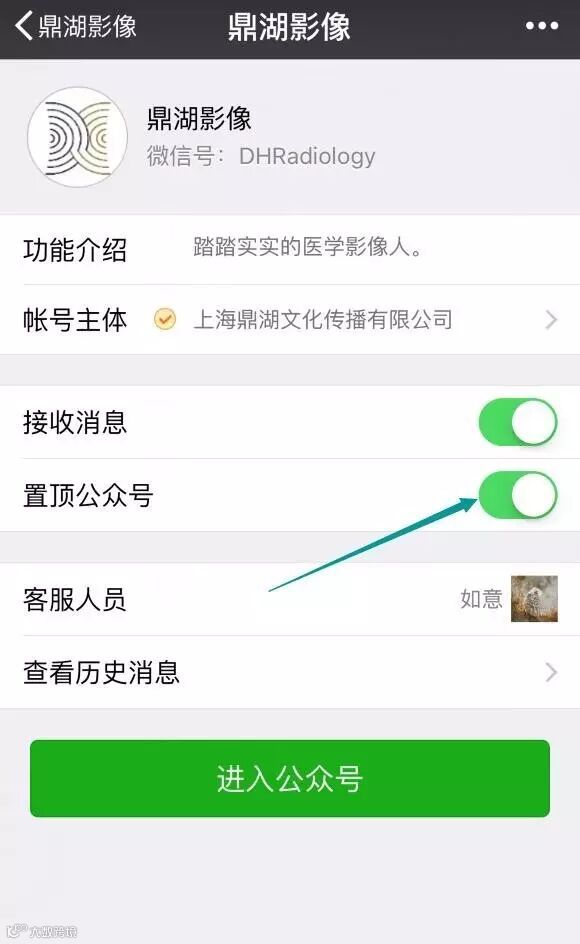
鼎湖影像还没置顶的,别睡了,起来置顶完再睡。这样你就不会错过一次次学习的机会,还可以赢得各种红包读片奖励、各类积分奖励。不要因为没有置顶错过而遗憾呦。
